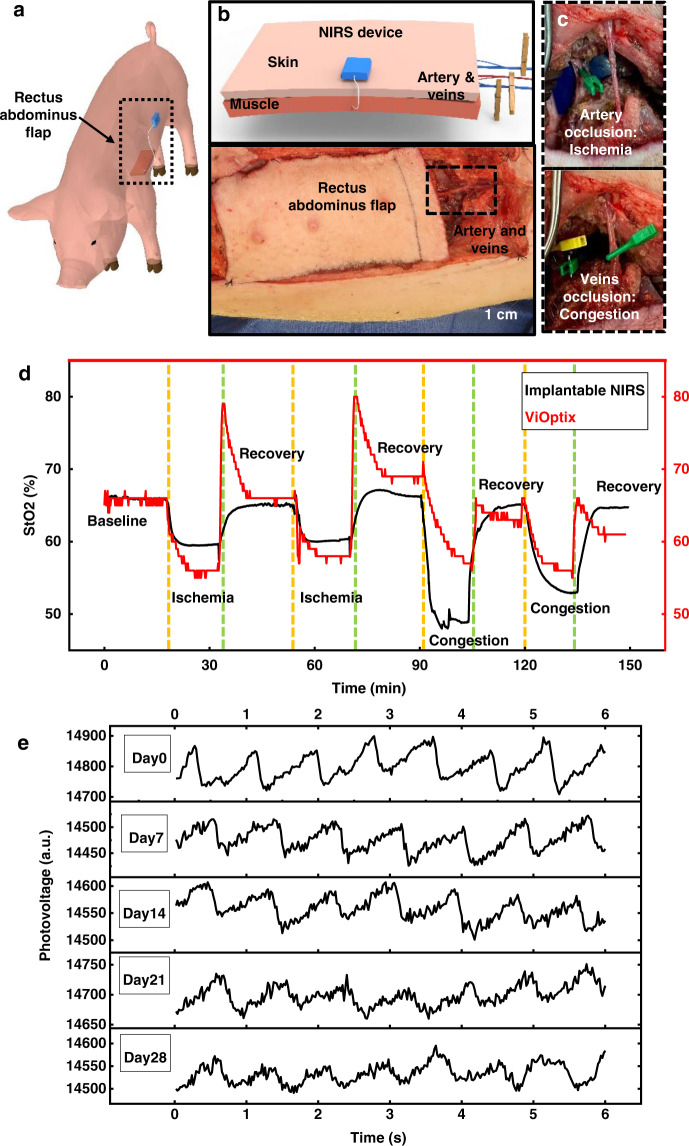
Fig. 4. In vivo measurements in a porcine flap model.
a, b Schematic illustrations of the implantation strategy using the left rectus abdominus flap in a porcine model. c Image of the occlusion of arteries and veins to simulate events of ischemia and congestion, respectively. d Measured tissue oxygenation saturation from the implantable NIRS probe and from a skin-mounted device (ViOptix) during multiple experimental cycles that simulate events of ischemia and congestion. e Measured patterns of pulsation recorded from the fingertips using a NIRS probe after immersion in phosphate buffered saline solution at 37 °C for various time periods. Source data are provided as a Source Data file.

