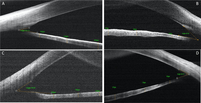
Fig. 1. Anterior chamber angle width and iris thickness in normal and PCG eyes.
Imaging of the anterior chamber angle with RTVue handheld anterior segment optical coherence tomography (RTVue HH-ASOCT), showing the anterior chamber angle width and iris thickness in the nasal and temporal quadrants in a normal infant (A, B) and in primary congenital glaucoma (C, D).

