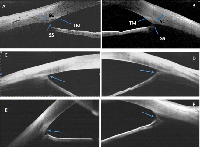
Fig. 2. Anterior chamber angle structures in normal and PCG infants.
Anterior chamber nasal and temporal angles as imaged by RTVue handheld anterior segment optical coherence tomography (RTVue HH-ASOCT) in a normal infant (A, B), versus in PCG (C, D, E, F). In normal infants, scleral spur (SS) could be detected, trabecular meshwork was identified (TM), and Schlemm’s canal (SC) was seen (A, B). Anteriorly inserted, thinned out iris, with severe trabeculodysgenesis and no identifiable angle structures as seen in a primary congenital glaucoma infant (blue arrow in C, D). An abnormal membrane clearly visualized extending from the iris to the trabecular meshwork (blue arrow in E, F), in another primary congenital glaucoma infant.

