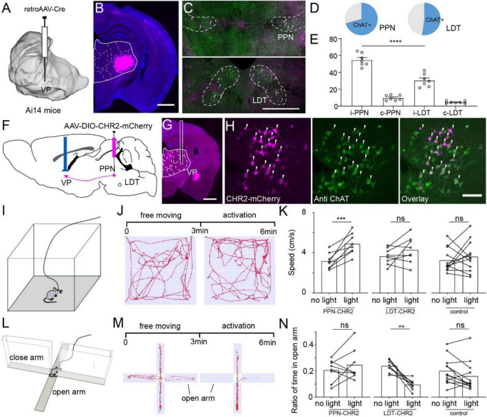
FIGURE 5.
Different roles of ChATPPN and ChATLDT projecting to VP. (A) Retro-AAV carrying Cre was performed in the VP of Ai14 reporter mice. (B) A typical coronal plane of an inject site. (C) Immunohistochemical staining of PPN or LDT neurons project to the VP. (D) Proportion of ChAT+ neurons projecting from PMT to VP. (E) Quantitative statistical proportion of ChAT+ neurons, originating from bilateral PPN and LDT. (F) Cre-dependent AAV virus expressing CHR2 and mCherry was injected in the PPN or LDT. (G) A typical coronal plane implanted with optical fiber. (H) Immunofluorescent staining of CHR2-labeled neurons. (I) A schematic diagram of mice in an open field. (J) The process of the experiment and typical motion trails of the mice moved in the open field. (K) Speed of the mice in the open field. (L) A schematic diagram of the mice in an elevated-plus maze. (M) The process of the experiment and typical motion trails of the mice moved in the elevated-plus maze. (N) The ratio of time that the mice spent in an open arm in the EPM. A scale bar, (B,G) 1,000 μm; (C) 500 μm; (H) 100 μm. Data are shown as mean ± SEM. (E) Two-sided t-tests, (K,N) paired t-tests, **p < 0.01, ***p < 0.001, ****p < 0.0001.

