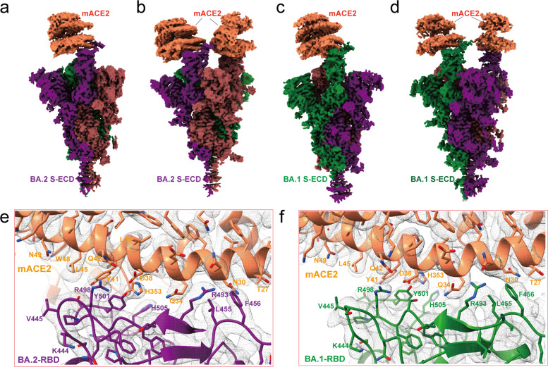
Fig. 5. Cryo-EM structure of the Omicron BA.2 and BA.1 spike trimers in complex with mACE2.
a, b Cryo-EM maps of the Omicron BA.2 spike protein-mACE2 complex with one RBD in “up” conformation at 3.2 Å resolution (a), and mACE2 complex with two RBDs in “up” conformation at 3.3 Å resolution (b), respectively. The three protomers are colored in purple, red and green, and the density for mACE2 is colored in coral. c, d Cryo-EM maps of the Omicron BA.1 spike protein-mACE2 complex with one RBD in “up” conformation at 3.1 Å resolution (c), and mACE2 complex with two RBD in “up” conformation at 3.2 Å resolution (d), respectively. The three protomers are colored in purple, red, and green, and the density for mACE2 is colored in coral. e, f Density maps and atomic models of the interaction interface in the BA.2 spike trimer-mACE2 (e) and BA.1-mACE2 complexes (f).

