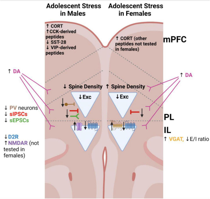
FIGURE 2.
Schematic of major changes to the medial prefrontal cortex identified in rodent models of adolescent stress. Triangles indicate pyramidal neurons. Arrows represent the direction of the alteration, relative to non-stressed controls of the same sex. CCK, cholecystokinin; CORT, corticosterone; D2R, dopamine receptor 2; D2R GPCR illustrated in blue; DA, dopamine; E/I ratio, excitatory/inhibitory balance; IL, infralimbic cortex; mPFC, medial prefrontal cortex (indicates alterations tested throughout this region); NMDAR, N-methyl-D-aspartate receptor; PL, prelimbic cortex; PV, parvalbumin-expressing; sIPSCs, spontaneous inhibitory postsynaptic currents; sEPSCs, spontaneous excitatory postsynaptic currents; SST-28, somatostatin-28 peptide; VGAT, vesicular GABA transporter 1; VIP, vasoactive intestinal peptide.

