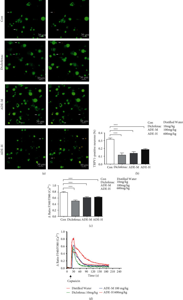
Figure 2.

Effect of ADE on the activity of TRPV1. (a) Representative Fura-2 ratio metric images of cultured DRG (L4–L6). Arrow indicates the DRG neurons in response to capsaicin. The color of the neurons switching from green to red indicates an increase in Ca2+ influx. (b) Percentage of DRG neurons responding to capsaicin in ipsilateral L4–L6 DRG neurons isolated from different group mice. (c) Activity of ipsilateral L4–L6 DRG neurons responding to capsaicin in neurons isolated from different group mice. (d) Representative traces illustrate that capsaicin elicited Ca2+ influx responses in ipsilateral L4–L6 DRG neurons. Each trace corresponds to the change in fluorescence ratio in a single neuron of cultured DRG neurons (∗p < 0.05, ∗∗p < 0.01, ∗∗∗p < 0.001∗∗∗p < 0.001; scale bar: 50 μm; n = 3).
