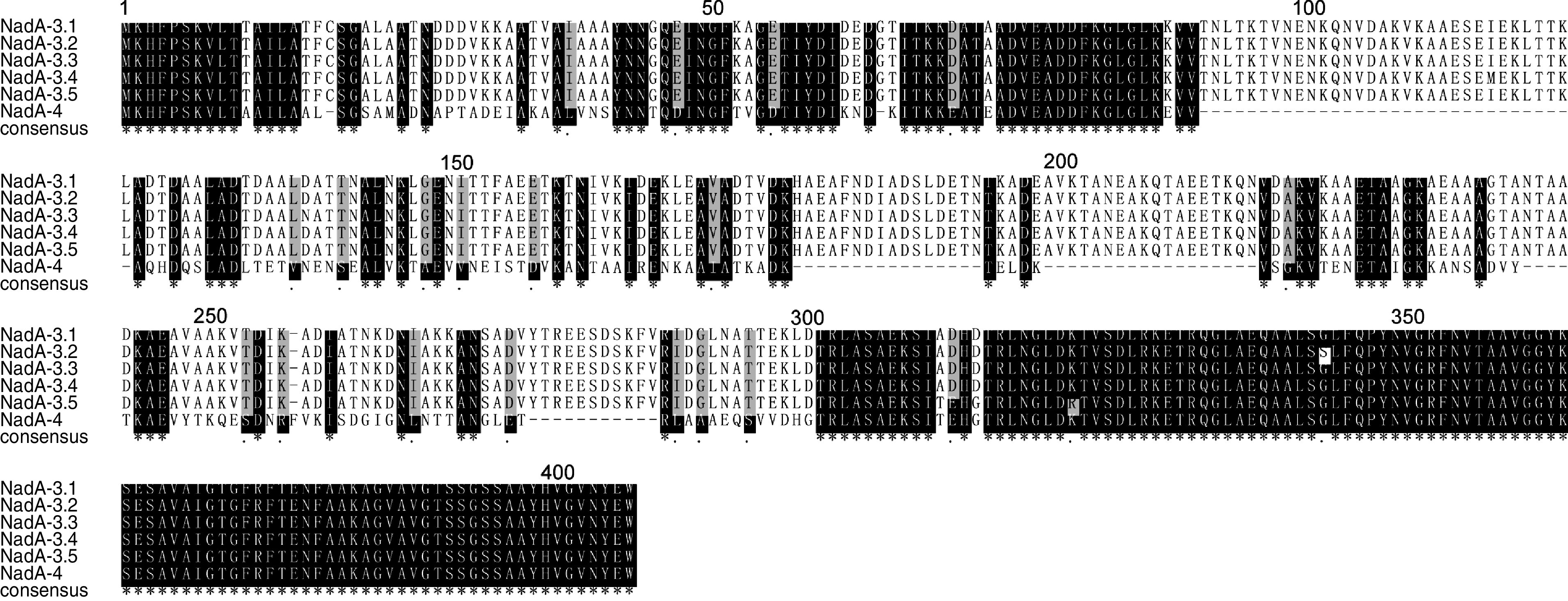
Fig. 3.

Alignment of six NadA subvariant sequences detected in this study. The designated identifications are shown for each sequence. Conserved residues are white, non-identical residues are black on white background, similar residues are grey and gaps are indicated by hyphens.
