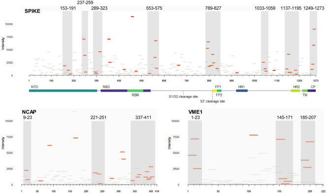
Figure 3.
Epitope mapping of SARS-CoV-2 spike, nucleoprotein, and membrane protein reveal distinct clusters of antibody reactivity. Differentially reactive peptides (adjusted p < 0.1, red tiles; adjusted p > 0.1, grey tiles) mapped on to their respective linear sequence. The x-axis of each panel represents the linear protein sequence arranged N-terminus (left) to C-terminus (right); y-axis represents the mean signal intensity for the COVID-19 convalescent sera (n = 22). Grey boxes demarcate epitope clusters, the sequence position is displayed for each. NCAP nucleoprotein, VME1 membrane protein, NTD N-terminal domain, RBD receptor-binding domain, RBM receptor-binding motif, FP fusion peptide, HR heptad repeat, TM transmembrane domain, CP cytoplasmic tail.

