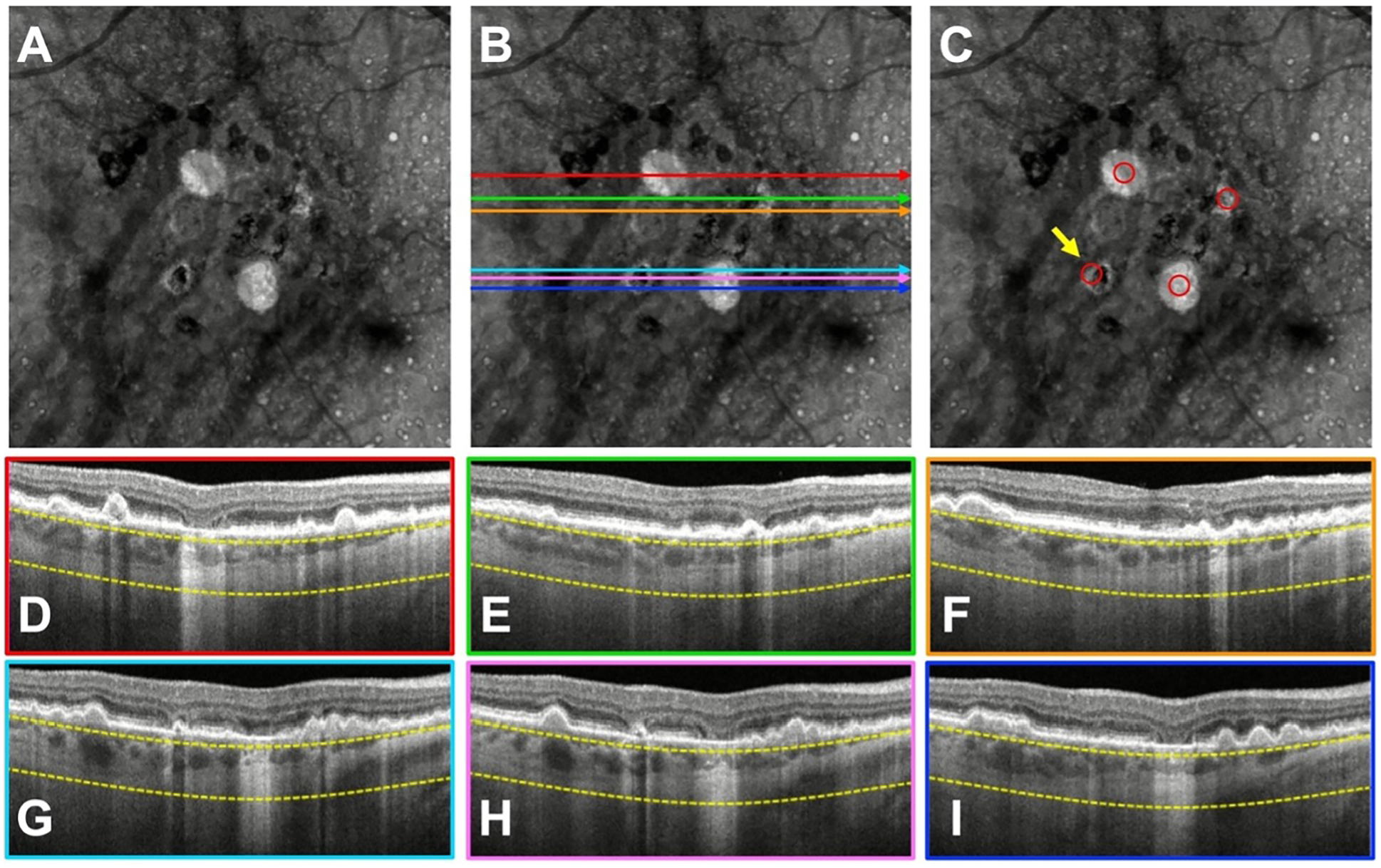
Figure 3.

A case with a donut lesion that was missed by three graders. A) Swept-source OCT (SS-OCT) en face structural image containing persistent choroidal hyper-transmission defects (hyperTDs). B, D–I) The same en face structural image shown in (A) with color-coded horizontal lines that correspond to the colored borders of the B-scans used by the graders to assess whether the region on the en face image corresponded to a true hyperTD. C) Structural en face image with the red circles identifying the hyperTDs. The yellow arrow is pointing to a hyperTD that three graders failed to correctly identify. As shown in the en face structural image with the red circles, the donut lesion has a greatest linear dimension (GLD) > 250 μm along the diagonal dimension. However, on the respective B-scans, the GLD of the horizontal hyperTDs are all < 250 μm.
