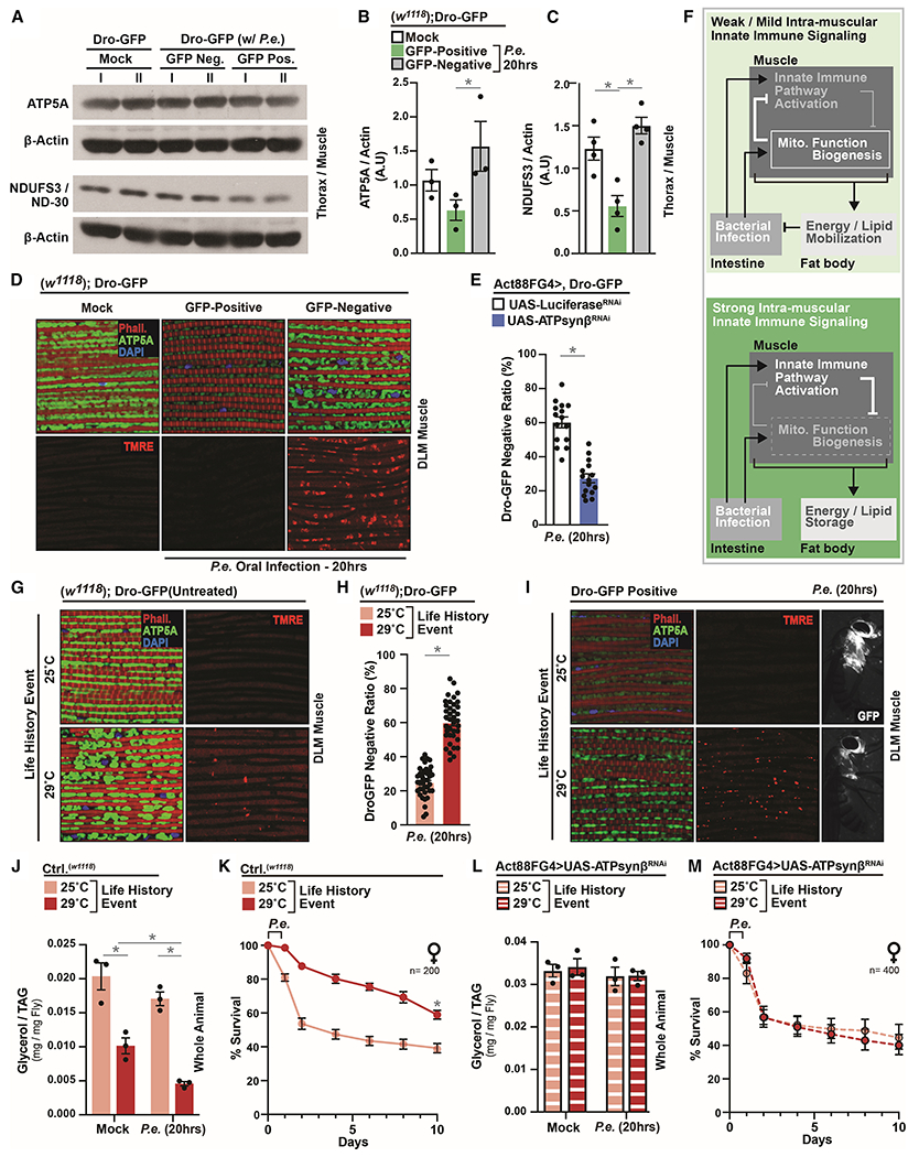
Figure 4: Life-history Events Influence Phenotypic Diversity of Host Immune-Metabolic Responses by Modulating Mitochondrial Dynamics and Host Energetic Trade-offs.

(A-D) Changes in mitochondrial dynamics in Dro-GFP flies after P.e. oral infection. (A) ATP5A and NDUF3/ND-30 protein levels (assayed by Western blot) in dissected thoraces/muscle; independent samples l and II are shown. (B-C) Quantification of three independent samples of (B) ATP5A and (C) NDUF3/ND-30, A.U.–arbitrary units. (D) Immunostaining to detect mitochondrial morphology (anti-ATP5A [green], F-actin filaments [Phalloidin, Red], and nuclei [DAPI, blue]) and TMRE fluorescent histochemistry (red).
(E) Ratios of Dro-GFP negative flies (percentage) within populations; n=300.
(F) Model summarizing conclusions.
(G-I) ATP5A immunostaining and TMRE fluorescent histochemistry (red) in dissected DLM muscle (G) before and (I) after infection; anti-ATP5A (green), F-actin filaments (Phalloidin, Red), and nuclei (DAPI, blue). (I) Right panel; corresponding induction of systemic drosocin (Dro-GFP). (H) Ratio (percentage) of Dro-GFP negative flies after P.e. oral infection; n=760.
(J) Total TAG levels of w1118 flies after P.e. oral infection; n=3.
(K) Survival of w1118 control flies after P.e. oral infection; n=200.
(L) Total TAG levels of Act88FGal4>UAS-ATPsynβRNAi flies after P.e. oral infection; n=3.
(M) Survival of Act88FGal4>UAS-ATPsynβRNAi flies after P.e. oral infection; n=400.
Error bars represent mean±SE, *P<0.01.
