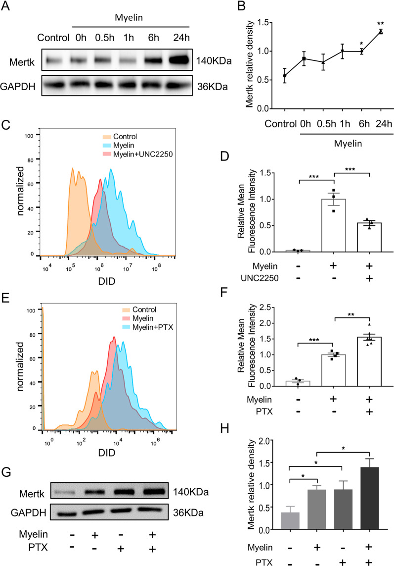
Fig. 4.
PTX upregulated Mertk expression and promoted phagocytosis in primary microglia exposed to myelin debris. A Representative immunoblots probed with antibodies against Mertk and GAPDH at different time points after myelin stimulation. B Quantification of Mertk bands normalized to GAPDH (n = 3 repeats per group). C, D Primary microglia were treated with UNC2250 (100 nM) for 2 h, and then con-cultured with myelin debris (0.01 mg/ml) stained with DID for 0.5 h, primary microglia were collected and myelin debris signal intensity in microglia were conduct by FACS (n = 3 repeats per group). E, F Primary microglia were treated with vehicle or PTX (25 μM) for 2 h, and then incubated with myelin debris (0.01 mg/ml) for 0.5 h. Myelin debris signal intensity in microglia were detected by FACS (n ≥ 3 repeats per group). G Representative immunoblots probed with antibodies against Mertk and GAPDH. H Quantification of the levels of Mertk normalized to GAPDH (n = 3 repeats per group). All data were presented as the mean ± SEM. *p < 0.05, **p < 0.01, ****p < 0.001

