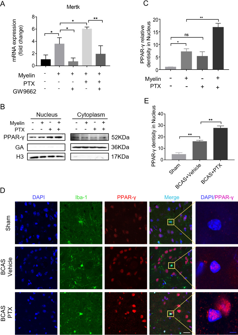
Fig. 5.
PTX upregulated Mertk by stimulating PPAR-γ nuclear translocation in vivo and in vitro. A Primary microglia were incubated with GW9662 (10 μM) for 1 h, followed with myelin debris (0.01 mg/ml) with or without PTX (25 μM) for 6 h. Quantitative RT-PCR analysis of Mertk mRNA in primary microglia (n = 3 repeats per group). B Representative immunoblots probed with antibodies against PPAR-γ, GADPH and H3 in nucleus and in cytoplasm of BV2 cells. C, Quantification of PPAR-γ levels normalized to H3 in nucleus (n = 3 repeats per group). D Immunofluorescent images of Iba-1 (green)/PPAR-γ (red)/DAPI (blue) colocalization in IC at Day 30 after BCAS. White scale bar: 20 μm, blue scale bar: 4 μm. E, Quantification of immunofluorescent intensity of PPAR-γ in DAPI area. The values were normalized to those of the control group (n = 4 mice per group). All data were presented as the mean ± SEM. *p < 0.05, **p < 0.01, “ns” means no significance (p > 0.05)

