Fig. 3.
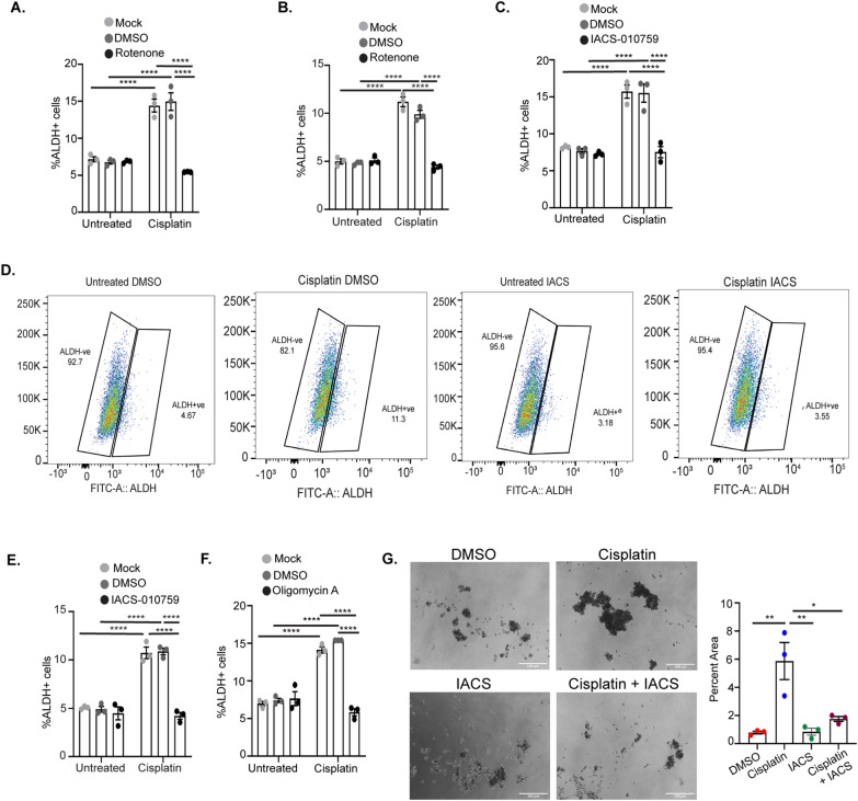
Mitochondrial OXPHOS inhibitors in combination with cisplatin block the platinum-induced increase in percent ALDH + cells. Percent ALDH + OVCAR5 (A) and OVSAHO cells (B) untreated or treated with 12 µM or 4 µM cisplatin, respectively, alone or in combination with DMSO or 5 µM Rotenone for 16 h followed by ALDEFLUOR assay. Percentage ALDH + OVCAR5 (C) and OVSAHO (D, E) cells treated with cisplatin alone or in combination with DMSO or 1 µM IACS-010759 for 16 h followed by ALDEFLUOR assay. D Shows gates used to determine ALDH + cells for one biological replicate of OVSAHO cells treated with DMSO or IACS in combination with cisplatin. F Percent ALDH + OVCAR5 cells treated with cisplatin alone or in combination with DMSO or 1 µM oligomycin for 16 h followed by ALDEFLUOR assay. Graphs display mean ± SEM percent ALDH + cells in N = 3 biological replicates. G Spheroid formation assay in OVCAR5 cells pre-treated for 3 h with DMSO or 1 μM IACS-010759 alone or in combination with 6 μM cisplatin and cultured for 14 days. For all untreated versus cisplatin treated, P values *< 0.05, **< 0.005, ***< 0.0005, ****< 0.0001

