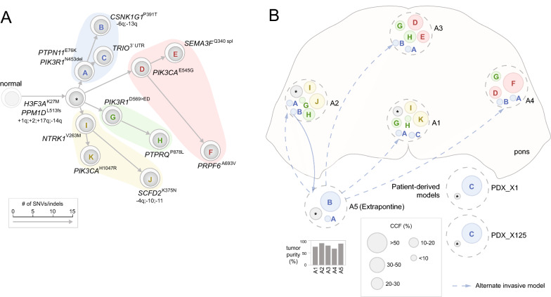
Fig. 5.
Extensive PI3K convergent evolution in patient 756. The two panels are drawn using the same style as Fig. 3. A Phylogenetic tree for patient 756 with four major branches each bearing a distinct PI3K mutation. B Spatial position and clonal composition of 5 profiled tumor regions including an extrapontine sample of unknown location (A5). Extrapontine invasions involving tumor cells from blue (clone A, B) are marked by blue arrows. Alternatively, the dotted blue arrows indicate that presence of clones A nd B in tumor regions within the pons could be caused by invasion from the extrapontine region A5. Two PDX samples (PDX_X1 and PDX_X125) derived from A1 shown a monoclonal lineage as only clone C was detected in both models

