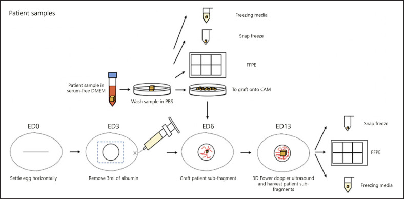
Fig. 2.
Overview of CAM protocol for patient samples. Fertilized eggs were collected from a local farm and designated as embryonic day (ED) 0. The eggs were mechanically wiped with dry paper towels and set horizontally in the Rcom Max 50 incubator at 37.5°C and 60% humidity. On ED3, 3 mL of the albumin was removed using a syringe and needle, and a small window of 1 cm2 was made in the center of the egg, and then sealed with a semipermeable adhesive film. On ED6, the patient tumor was collected in serum-free DMEM, washed in PBS, and cut into fragments of approximately 3 × 3 × 3 mm to be grafted onto the chorioallantoic membrane (CAM). The remaining fragments of the patient tumor was kept in freezing media, snap frozen and fixed in formalin before being embedded in paraffin. The percentage vascularity and volume of the grafted patient tumor was then measured using the 3D power Doppler ultrasound and was allowed to grow till ED13, before comparing the differences in vascularity and volume. The grafted patient tumor was then divided into subfragments to be kept in freezing media, snap frozen or fixed in formalin and embedded in paraffin. Comparisons in the FFPE samples before and after grafting into the CAM could be made.

