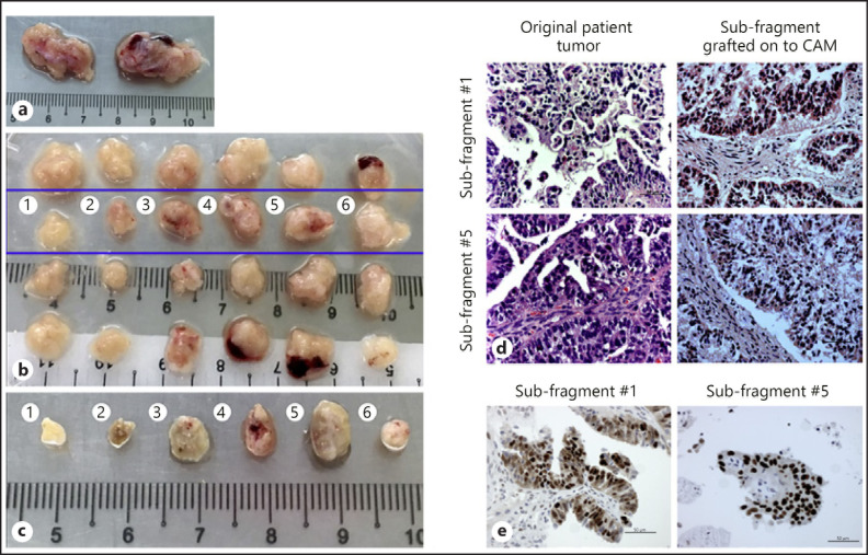
Fig. 3.
Engraftment of ovarian cancer patient fragments onto chick CAM. a Original patient tumor sample collected from the operating theater. b Each tumor sample was cut into 12 smaller fragments (as described in Fig. 2). The top row was kept in freezing media for future experiments, second row was grafted onto the CAM on ED6, subfragments of the third row were FFPE-treated and the bottom row was snap frozen in liquid nitrogen. Subfragments 1–6 in the second row were grafted onto the CAM on ED6. c Photograph of harvested tumor subfragments from the CAM on ED13. The subfragments from the original tumors from the blue box (b) were grafted onto the CAM on ED6 and left till ED13 before they were scanned by 3D ultrasounds and excised from the CAM. The photograph depicts the tumor subfragments excised from the CAM. The tumors harvested were than fixed in 10% formalin and embedded in paraffin before H&E staining was performed by the Department of Pathology in National University Hospital of Singapore. d H&E staining of original ovarian cancer patient fragment 1 (top left) and 5 (bottom left) and subfragment 1 (top right) and 5 (bottom right) after grafting onto CAM for 7 embryonic days. The subfragments post-grafted onto the CAM maintained similar morphology and histology compared to the original ovarian cancer patient fragment. Scale bars, 50 µm. ×40. e Immunoexpression of p53 in original patient fragment 5 (left) and subfragment 5 (right) post-grafting onto CAM. The subfragments post-grafted onto the CAM maintained similar immunoexpression of p53 compared to the original ovarian cancer patient fragment. Scale bars, 50 µm. ×40.

