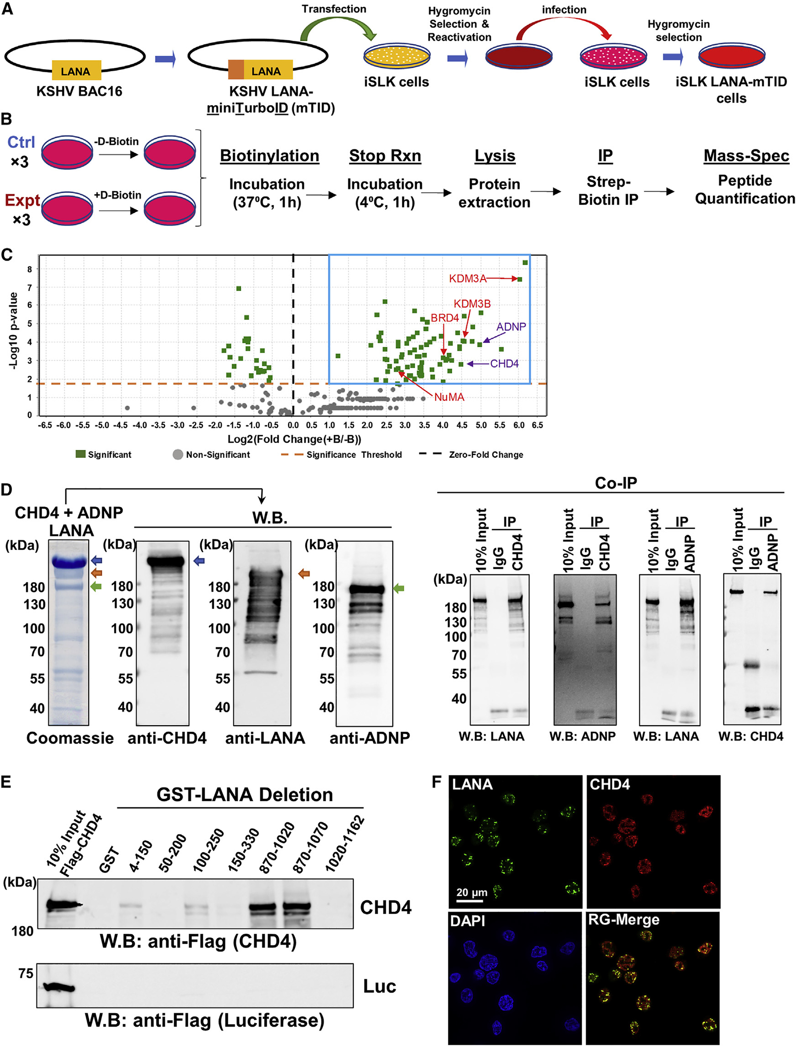
Figure 2. LANA interacts with the CHD4 and ADNP.

(A) A schematic diagram of preparation of recombinant KSHV-infected iSLK cells.
(B) Experimental design for preparing samples for protein ID. iSLK-LANA mTID cells were left unincubated (−) or incubated (+) with D-biotin (500 μM) for 1 h. Unincubated (−) cells were used as control samples.
(C) The volcano plot represents proteins identified in close proximity to LANA. Proteins with an abundance Log2 FC of greater than or equal to 1 and p value less than 0.05 were selected and are shown by blue box. Log2 FC was calculated as (+B/−B) where + B and −B indicate presence and absence of D-biotin, respectively. The t test was used for calculating the p value. Purple color, ChAHP components; Red color, previously known LANA interacting proteins.
(D) LANA, CHD4, and ADNP complex, which consists of Flag-LANA (gold arrow), Flag-CHD4 (blue arrow), and His-ADNP (green arrow), was prepared by co-infected three recombinant baculoviruses, and the protein complex was isolated with FLAG-affinity purification. The authenticities of the respective protein bands were confirmed by immunoblotting with specific antibodies. Coomassie staining is shown. Ten percent of the reaction before immunoprecipitation was used as controls. W.B., western blotting; CoIP, Co-immunoprecipitation.
(E) An equal amount (1 μg) of each LANA deletion protein purified from E. coli was incubated with full-length Flag-tagged luciferase (1 μg) or Flag-tagged CHD4 (1 μg) in binding buffer and interaction was probed with anti-Flag antibody. LANA deletion proteins are presented in Figure S2C.
(F) CHD4 and LANA were probed with anti-CHD4 rabbit monoclonal antibody and anti-LANA rat monoclonal antibody, respectively. Images were acquired with Keyence fluorescence microscopy. Scale bar, 20 μm. (D–F) n = 3 biological replicates, and one representative is shown. (C) Each protein ID was performed with three biological replicates.
