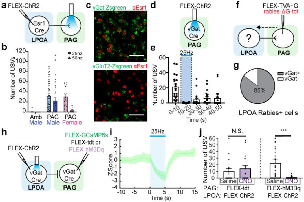
Figure 2. LPOAvGat -> PAGvGat -> PAGvGluT2 di-synaptic disinhibition circuit triggers USVs.

a-b, LPOAEsr1/ChR2 axon terminal photostimulation in the PAG evokes USV calls. a) experimental design, b) number of USVs detected in the Amb (Males, N=5 animals) and the PAG (Males N=13 animals, Females N=4 animals) Mean ± s.e.m. c, sample images of LPOAEsr1+ co-expression with vGat-Zsgreen (upper, N=4 animals, 3 sections/animal counted) and vGluT2-Zsgreen (lower, N=3 animals, 3 sections/animal counted). Scale bar = 50μm. d-e, photostimulation (blue bar) of male inhibitory PAGvGat/ChR2 neurons during social interaction with an awake female blocks natural USVs. d) experimental design to express ChR2 in local inhibitory PAG neurons and e) quantification of evoked USV syllables. Mean ± s.e.m. N=3 animals, 19 trials. f, experimental design to assess direct anatomy to LPOA by retrograde tracing from PAGvGat cells. g, percentage of tdt expression (rabies) in LPOA vGat+ cells. N=3 animals, 320 cells counted. h, experimental design to express ChR2 in LPOAvGat and GCaMP6s or tdt or hM3Dq in PAGvGat. i, mean z-score of fiber photometry signal from PAGvGat/GCaMP with 25Hz photostimulation (blue bar) on LPOAvGat/ChR2 cells. Solid line indicates mean of all trials; shaded region indicates 95% confidence interval. N=4 animals, 4 trials/animal. j, USVs are silenced during LPOAvGat/ChR2 photostimulation and PAGvGat/hM3Dq CNO. Mean ± s.e.m. N=3 animals, 36 trials (hM3Dq), 18 trials (tdt control). Paired Wilcoxon test, two sided, ***=0.005.
