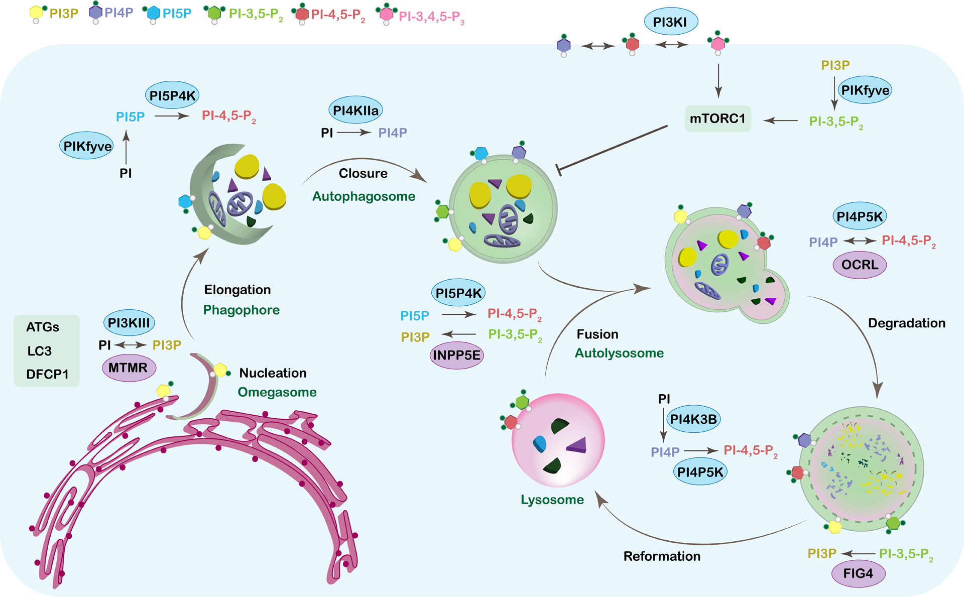
Figure 2. Phosphoinositides in autophagy.

Autophagy is a highly regulated multistep process that is essential for recycling cytosolic components. Too much or too little autophagy leads to severe human diseases from neurodegeneration to cancer. Phosphoinositides play key roles in every step of this process from nucleation and the biogenesis of the phagophore to the reformation of the lysosome. Distinct pools of phospholipids characterize each membrane of the autophagic organelles. This phospholipid identity is ensured by specific kinases and phosphatases and their binding partners. The figure shows all the individual steps of autophagy starting with the nucleation and ending with the reformation of the lysosome. The corresponding phosphoinositide species that are known to play a role in regulating each step are shown on the membrane of the structure they are associated with and are color-coded. The kinases (blue oval) and phosphatases (purple oval) that are associated with regulating the phosphoinositide species at each step are also included. For simplicity, this model does not show all known binding partners, but these are discussed within the text.
