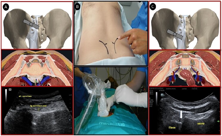
Fig. 1.
Technique used for US-guided injection into the SI joint
(A) Anatomical and US image when approaching the upper two-thirds of the SI joint (syndesmosis). (B) Positioning of the patient and approach in the joint plane with a curved transducer. (C) Anatomical and US image when approaching the lower one-third of the SI joint (synovial).

