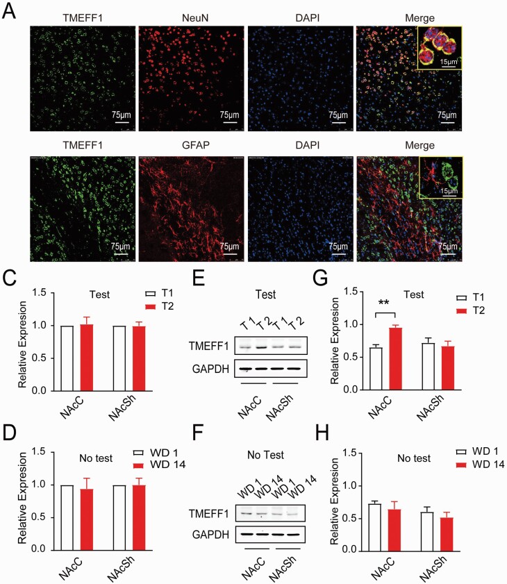
Figure 3.
Expression of TMEFF1 in response to incubation of morphine craving. (A, B) The subcellular localization of TMEFF1 by using the laser scanning microscope. The TMEFF1 protein labeled with green fluorescence, A: neuron nuclei (NeuN), and B: astrocytes (GFAP) labeled with red fluorescence (Scale bar = 75 μm). (C) The expression of TMEFF1 mRNA between T1 and T2 in NAcC or NAcSh. (D, E) Western-blot analysis of TMEFF1 and GAPDH expression between T1 and T2 in NAcC or NAcSh. **P < .01, vs T1 in NAcC. (F) The expression of TMEFF1 mRNA between WD1 and WD2 in NAcC or NAcSh without test. (G, H) Western-blot analysis of TMEFF1 and GAPDH expression between WD1 and WD2 in NAcC or NAcSh without the test.

