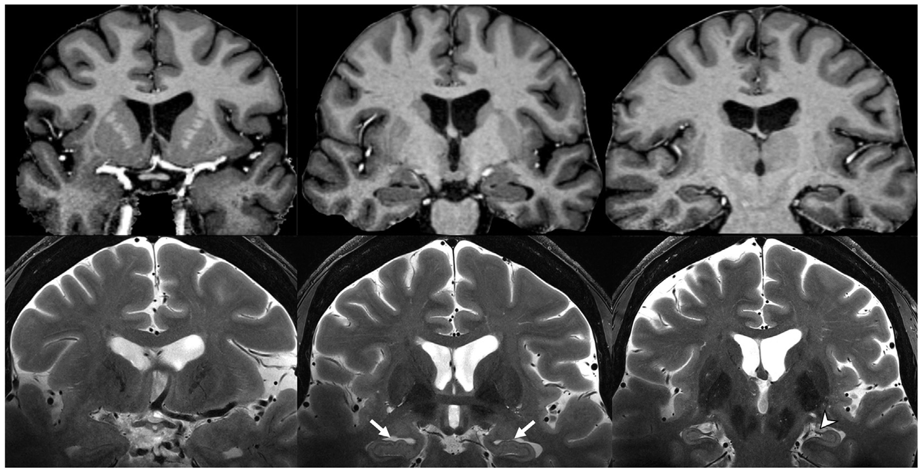
Fig. 1.

Skull-stripped coronal plane T1 MP2RAGE (top row) and unprocessed T2-weighted (bottom row) MR images. Note the contrast between grey and white matter on the T1 MP2RAGE, and the hippocampal digitations (white arrows), fimbria (arrowhead), and stratum layers of the hippocampus resolved on the T2-weighted images at 7-T.
(MP2RAGE, magnetization prepared, two rapid-acquisition gradient-echo).
