Fig. 2.

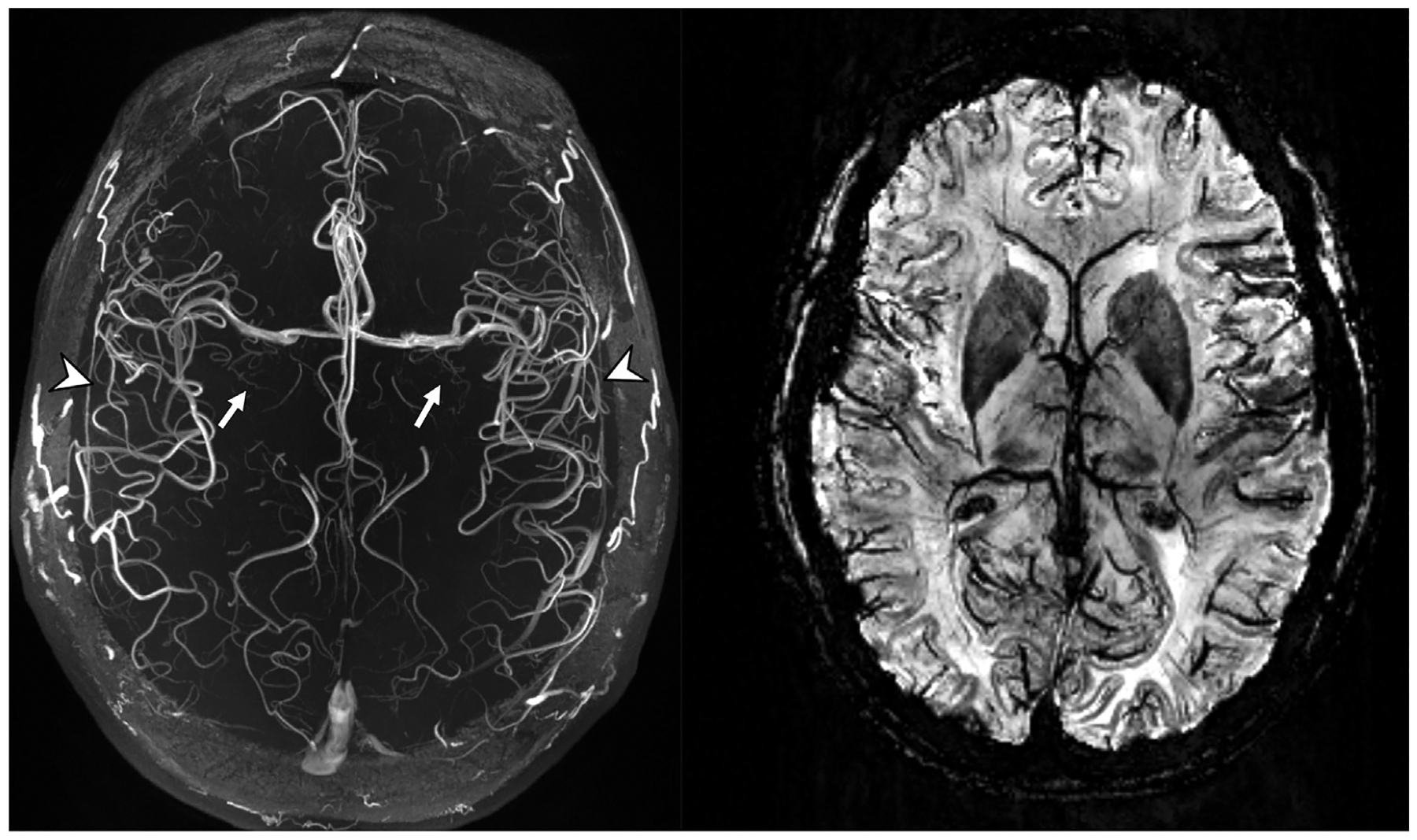
Axial plane TOF-MRA MIP (left) and SWI MinIP (right) MR images. Note the conspicuity of the lenticulostriate arteries (white arrows) arising from the MCAs, the high contrast seen in the insular branches (arrowheads) of the MCAs, and the high susceptibility and spatial resolution allowing better definition of deoxyhemoglobin in the smaller veins on the SWI image.
(MCA, middle cerebral artery; MinIP, minimum intensity projection; MIP, maximum intensity projection; SWI, susceptibility-weighted imaging; TOF-MRA, time-of-flight, magnetic resonance angio-graphy).
