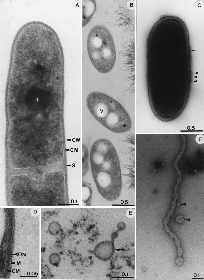
FIG. 2.
Electron micrographs of strain JF-5 grown in aerobic TSB medium lacking Fe(III) (A) or in Fe-TSB medium (B to F). (A, B, and D to F) Thin-section micrographs. (C) Whole cell, negatively stained with 2% phosphotungstate. Single blebs (arrowheads) and a short “chain” consisting of four blebs (small arrow) are visible on the cell periphery. (D) Cell envelope of JF-5 with features of a gram-negative cell. (E) Micrograph showing that blebs and extrusions are membrane surrounded (arrow). (F) The long extrusion which originates from the cytoplasmic membrane reveals constrictions. Arrowheads indicate single free blebs of different sizes. Bar lengths are shown in micrometers. Abbreviations: CM, cytoplasmic membrane; CW, cell wall; I, inclusion body; S, septum (division point); M, murein layer; OM, outer membrane; V, vesicle.

