. 2022 May 31;11:e69802. doi: 10.7554/eLife.69802

Figure 3. HIIT regulates proteins involved in skeletal muscle calcium sensitivity and handling HIIT did not alter the abundance of myosin heavy chain (MYH1: MyHC2x, MYH2: MyHC2a, MYH4: MyHC2x, MYH7: MyHCβ; A) or light chain (B) isoforms.

(C). HIIT regulates proteins controlling myosin phosphorylation. (D). HIIT reduces abundance of subunits of the dihydropyridine receptor. (E). Summed total protein abundances display downregulation of the dihydropyridine receptor. (F). HIIT does not alter the abundance of ryanodine receptor 1. Summary statistics are mean ± SEM (n=8). ^ FDR <0.05. * p<0.05, ** p<0.01, *** p<0.001.

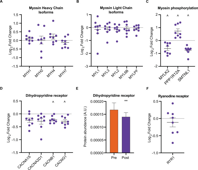
Figure 3.

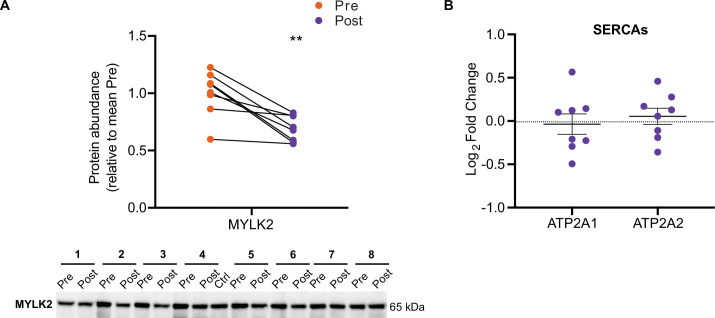
Figure 3—figure supplement 1. HIIT decreases MYLK2 abundance.

Figure 3—figure supplement 1.

(A) Immunoblotting analysis confirms the downregulation of MYLK2. Representative images confirming equal loading are displayed in Figure 5—figure supplement 2. (B). No change in the abundance of sarcoplasmic/endoplasmic reticulum calcium ATPases 1–3 (SERCAs). Summary statistics are mean ± SEM (n=8). * p<0.05, ** p<0.01, *** p<0.001.
Figure 3—figure supplement 1—source data 1. Full image and annotation of MYLK2 immunoblot.
Figure 3—figure supplement 1—source data 2. Raw ImageLab file of MYLK2 immunoblot.
Figure 3—figure supplement 1—source data 3. Raw quantification data of MYLK2 immunoblot.