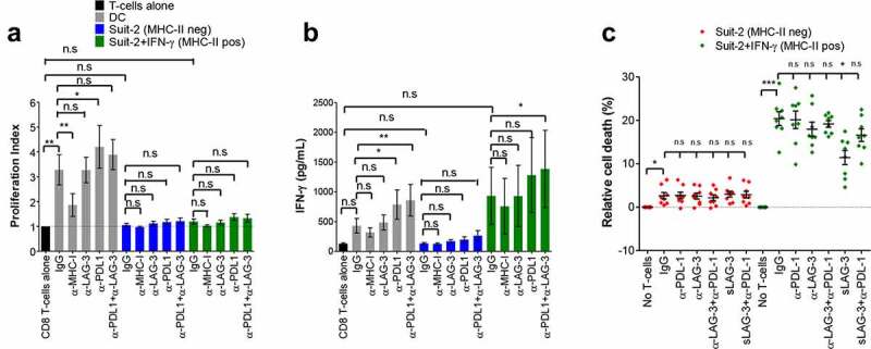
Figure 2.

MHC-II/LAG-3 axis contributes to CD8+ T-cells cytotoxic function. Allogenic activated CD8+ T-cells from healthy donors were cultured with MMC-treated PDAC cells (T-cell:tumor ratio 2:1) in the presence of blocking antibodies as indicated or isotype control (IgG) for 4 days. T-cell proliferation was determined by CFSE staining and results are expressed as mean values and S.E.M of the proliferation index (A) determined as described in Materials and Methods. The supernatants of the co-cultures were collected and assayed for IFN-γ by ELISA (B). Allogenic mature DCs (T-cell:DC ratio 10:1) were used as positive control in these experiments. For determination of cytotoxic activity of CD8+ T-cells, activated CD8+ T-cells from HLA-A2-negative donors were incubated overnight with untreated or IFN-γ-treated HLA-A2-positive Suit-2 cells. Where indicated, blocking antibodies, isotype control or sLAG-3 was added. Cell viability of tumor cells in these co-cultures was determined by staining with EthD-1 and calcein-AM. Tumor cells gated on HLA-A2+ that were negative for EthD-1 and positive for calcein-AM were considered viable cells (C). All data were analyzed by one-way ANOVA. *P < 0.05; **p < 0.01; ***p < 0.001; n.s: non-significant. Values shown in A and B were from independent experiments with 3 donors; in C is shown values from 4 donors performed in duplicate.
