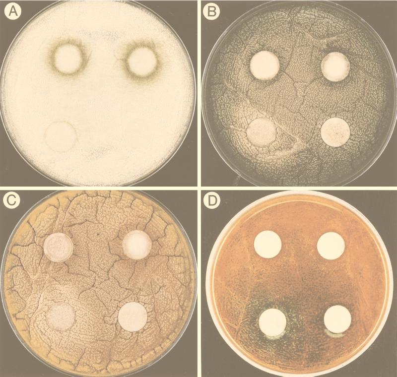
FIG. 3.
Photographs showing the effect of linoleic acid on Aspergillus development. The pictures were taken 40 h after inoculation. In each photograph the lower right disc was the solvent control, the lower left disc contained 0.1 mg of linoleic acid, the upper left disc contained 0.5 mg of linoleic acid, and the upper right disc contained 1 mg of linoleic acid. (A) Precocious and increased conidiation in A. flavus 12S with increasing amounts of linoleic acid. (B) The 0.1-mg linoleic acid treatment induced sexual development (visualized by a halo of yellow Hülle cells) in A. nidulans FGSC4, whereas the 0.5- and 1-mg treatments induced asexual development (visualized by the production of green conidia). (C) The 0.1-mg linoleic acid treatment induced sexual development (visualized by a halo of yellow Hülle cells) in A. nidulans WIM126, whereas the 0.5- and 1-mg treatments induced asexual development (visualized by the production of brown conidia). (D) Plate shown in panel C after treatment with the laccase substrate that stains sexual primordia green.

