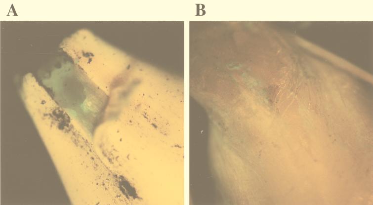
FIG. 4.
Photographs of the embryo areas of germinating barley seeds obtained with the epifluorescence stereomicroscope. (A) Green fluorescent areas corresponding to bacterial cells near the embryo under the glume. (B) Green fluorescent spots corresponding to bacterial cells near the point of root emergence observed after the glume was peeled off. Magnification range, ×10 to ×40.

