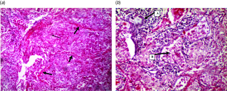
Fig. 2.
[colour online]. Histological examination of elephant lung. (a) The thick arrows indicate fibrous encapsulating material. The thin arrow indicates the characteristic stellate formation of a tubercle. Note the lost architectural pattern of the lung due to collapsed alveoli and the increased mononuclear infiltration. (b) Arrow (A) shows a giant cell in the midst of other inflammatory cells while arrow (B) shows cellular debris in an alveolar (H&E stain, ×100).

