Figure 1.
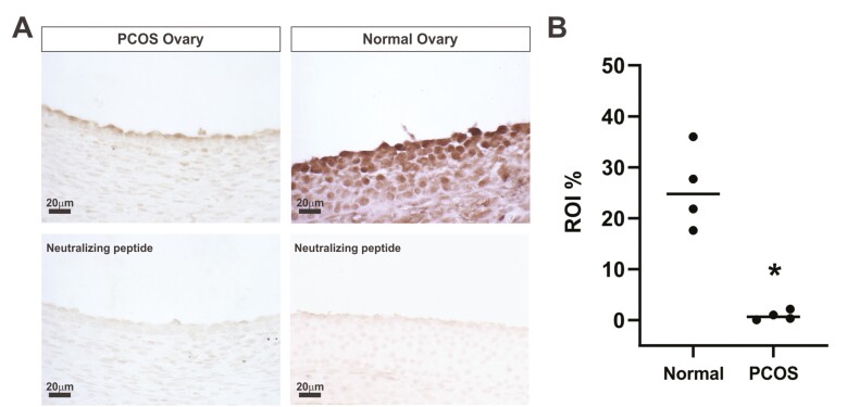
A, ZNF217 immunohistochemical staining of normal ovarian tissue (right) and polycystic ovary syndrome (PCOS) ovarian tissue (left). Images were taken at 40× magnification. In comparison to the theca cells of the normal cycling ovary, ZNF217 protein expression was decreased in theca layer of follicles of PCOS ovarian specimens. Antibody neutralized with the ZNF217 immunogenic peptide was used in the bottom 2 images to confirm specificity of the immunoperoxidase signal. B, Comparison of ZNF217 staining intensity in the designated region of interest (ROI, theca interna) revealed a significant decrease in the theca cell layer of PCOS ovarian tissue (N = 4) compared to normal ovarian tissue (N = 4); *P < .001.

