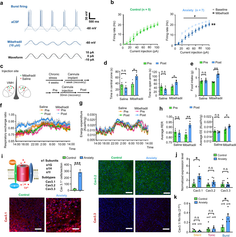
Fig. 4. T-VGCC mediated enhancement of burst firing in dmVMH neurons under chronic stress.
a Evoked burst firing trace of dmVMH neurons without and with T-VGCC antagonist (mibefradil, 10 μM), 10 pA current injection was given in cosine waveform. b Effects of mibefradil on suprathreshold activity in dmVMH burst firing neurons in control (n = 5 cells from 4 mice) and anxiety groups (n = 7 cells from 5 mice). Mibefradil inhibited T-VGCC and caused a rightward frequency-current curve shift (two-way ANOVA, control, P = 0.2140, F (1, 8) = 1.854; anxiety, P = 0.0077, F (1, 12) = 10.22, with Bonferroni correction). c Schematic of cannula implanted sites and experimental strategy of T-VGCC blockage in the VMH of chronic stress-treated mice. d Behavioral test before and after delivery of mibefradil or saline; left, residence time in central area of open field increased after mibefradil application (n = 4 in each group, P = 0.0142, paired Student’s t-test); right, residence time in open arm increased after mibefradil application (n = 4 in each group, P = 0.0228, paired Student’s t-test). e–h Administration of mibefradil in stressed mice increased food intake (n = 4 in each group, P = 0.0030, unpaired Student’s t-test), average RER (P = 0.00671, paired Student’s t-test) and average EE (P = 0.0482, paired Student’s t-test). RER and EE curve shifted (two-way ANOVA, RER: P = 0.0067, F (1, 6) = 16.42; EE: P = 0.0301, F (1, 6) = 7.993, with Bonferroni correction) after applying mibefradil. No comparable changes were observed in saline group. i Schematic Structure of voltage-gated calcium channel located on cell membrane (left top). Representative immunofluorescence showing Cav 3.1 (left bottom), Cav 3.2 (right top), and Cav 3.3 (right bottom) expression in dmVMH of control and anxiety mice respectively, significantly increased Cav3.1 expression observed after chronic stress (Cav3.1+ cells counting, unpaired Student’s t-test, P < 0.001). Scale bar: 100 μm. j Quantification of T-VGCCs expression in dmVMH tissue between control (n = 5 mice) and anxiety groups (n = 6 mice). Expression of Cav 3.1 was significantly upregulated under chronic stress conditions (unpaired Student’s t-test, P = 0.0232). k Single-cell qRT-PCR analysis of T-VGCC expression in dmVMH neuronal subtypes between control (n = 16 cells) and anxiety groups (n = 13 cells). Expression of Cav 3.1 in burst firing neurons was significantly upregulated in anxiety group (unpaired Student’s t-test, Cav3.1, P = 0.0164). means ± SEM. *P < 0.05, **P < 0.01.

