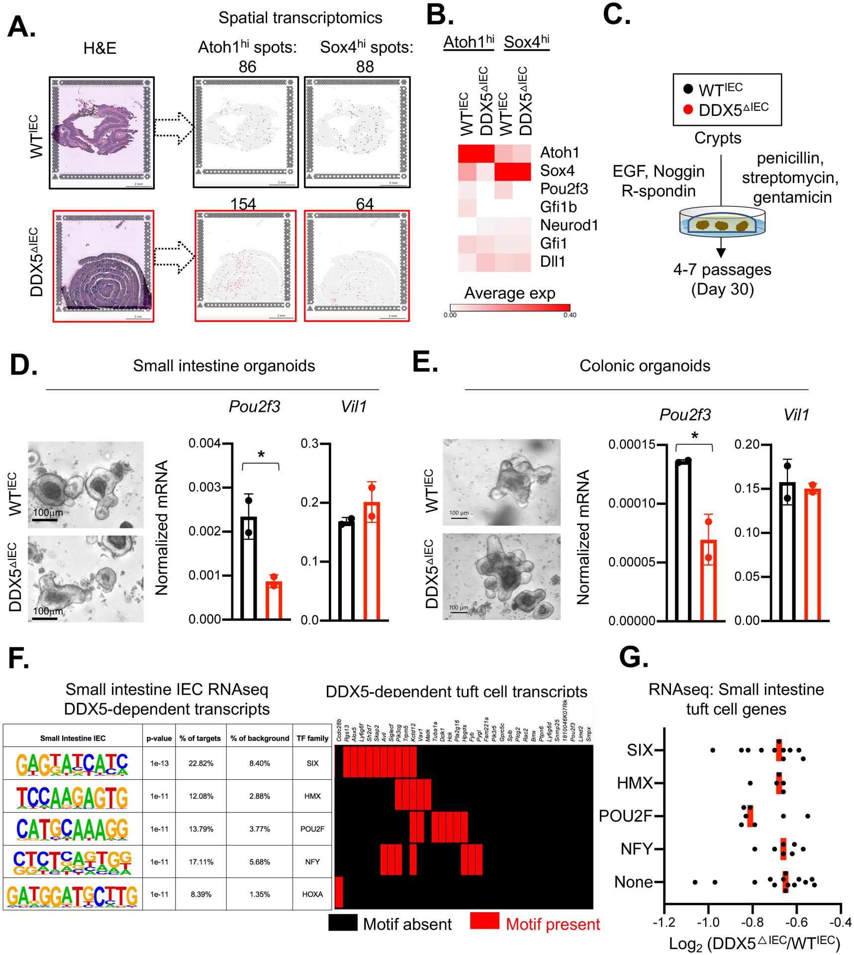
Figure 2. Spatial transcriptomic analysis revealed altered secretory lineage progenitor signature in the DDX5ΔIEC mice.

A. Left: H&E staining of the small intestinal swiss rolls from one pair of female WTIEC and DDX5ΔIEC littermates used for the spatial transcriptomic analysis (Visium, 10X Genomic). Right: Loupe browser displaying Atoh1high or Sox4high spots on each tissue section. Scale bar: 2mm.
B. Heatmap indicating the average expression of IEC lineage-specifying genes in Atoh1high or Sox4high spots indicated in A.
C. Schematic of establishing intestinal crypt organoid cultures.
D. Left: Representative bright field images of organoids established from small intestinal crypts of WTIEC and DDX5ΔIEC mice following the protocol outlined in50. Scale bar, 100μm. Right: Normalized mRNA expressions of Pou2f3 and Vil1 from WTIEC (n=2) and DDX5ΔIEC (n=2) small intestinal organoids cultured over 4–7 passages. Each dot represents the result from an independent organoid experiment. Results were averaged from two independent biological experiments. * p<0.05 (unpaired t-test).
E. Left: Representative bright field images of organoids established from colonic crypts of WTIEC and DDX5ΔIEC mice following the protocol outlined in50. Scale bar, 100μm. Right: Normalized mRNA expressions of Pou2f3 and Vil1 from WTIEC and DDX5ΔIEC colonic organoids cultured over 4–7 passages. Each dot represents the result from an independent organoid experiment. Results were averaged from two independent biological experiments. * p<0.05 (unpaired t-test).
F. Left: HOMER de novo analysis identified the top 5 motifs present at the promoters (defined as −1 kb to +500 bp from the transcription start site) of all ileal IEC DDX5-dependent genes identified in Figure 1A39. Promoters from all other IEC expressed genes were used as background. Right: The presence (red) or absence (black) of the motifs indicated on the left at the tuft cell DDX5-dependent gene promoters.
G. Average log2 fold change of the transcript abundance of tuft cell-expressed genes harboring SIX, HMX, POU2F, NFY motifs, or None of the above motifs at their promoters in ileal IECs from two independent pairs of WTIEC and DDX5ΔIEC littermates. Each dot represents one gene.
