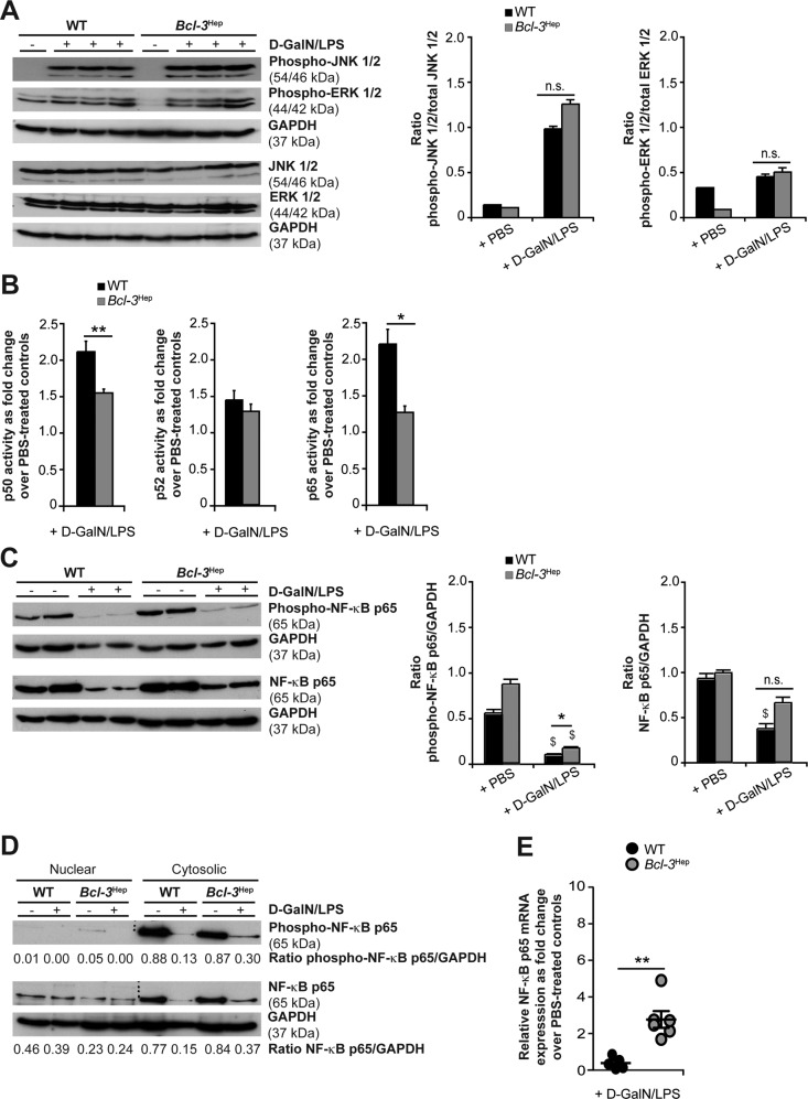
Fig. 3. Activation of hepatic JNK, ERK and NF-κB in Bcl-3Hep and WT mice in response to d-GalN/LPS.
A Immunodetection of phospho-JNK (Thr183/Tyr185), phospho-ERK (Thr202/Tyr204) and total JNK/ERK protein in whole liver tissue lysates from Bcl-3Hep and WT mice at 4 h post d-GalN/LPS challenge. B NF-κB p65, p50 and p52 activity was quantified by a functional binding assay. Ratio of relative NF-κB p65/p50/p52 activity in d-GalN/LPS-challenged Bcl-3Hep vs. WT mice is shown (means of n = 7 WT + d-GalN/LPS, n = 8 Bcl-3Hep + d-GalN/LPS and n = 7 PBS-treated controls per genotype ± SEM). C Immunoblotting of phospho-NF-κB p65 (Ser536) and total NF-κB p65 in whole liver tissue lysates and D after cytosolic vs. nuclear cell fractionation. E Relative hepatic NF-κB p65 gene expression in d-GalN/LPS-challenged Bcl-3Hep vs. WT mice as fold change over PBS-treated controls (means of n = 6 mice per genotype ± SEM). In A, C and D representative immunoblots with densitometric analysis are shown. *p < 0.05, **p < 0.01 for WT vs. Bcl-3Hep and $p < 0.05 for PBS vs. d-GalN/LPS using unpaired, two-tailed Student’s t-test (A–C) or Mann–Whitney U test (E).

