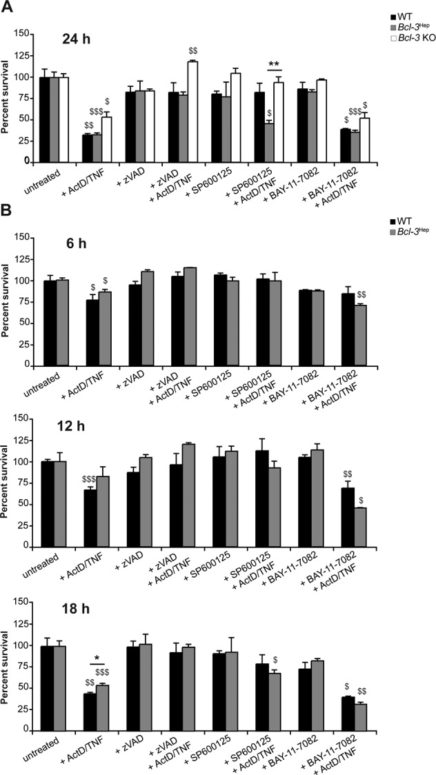
Fig. 4. Rate of TNF-induced hepatocyte cell death in vitro.

A Primary hepatocytes derived from Bcl-3Hep, WT, and Bcl-3 KO mice were treated ex vivo with ActD (200 ng/mL) and TNF (10 ng/mL) to induce TNF-R driven cell death. Pan caspase inhibitor zVAD (50 µM), JNK inhibitor SP600125 (100 µM), or IKK inhibitor BAY-11-7082 (10 µM), where ever indicated, were added 1 h before ActD/TNF-treatment to examine the activity of caspases, JNK and NF-κB signaling in TNF-induced cell death in Bcl-3Hep, WT, and Bcl-3 KO hepatocytes. After 24 h cell viability was assessed by MTT colorimetric assay relative to untreated samples. B Time course analysis of ActD/TNF-induced cell death in Bcl-3Hep and WT hepatocytes determined by MTT assays after 6, 12, and 18 h. Numerical data in mean ± SEM of A three or B two independent experiments performed at least in duplicate readings. *p < 0.05, **p < 0.01 for WT vs. Bcl-3Hep or Bcl-3Hep vs. Bcl-3 KO and $p < 0.05, $$p < 0.01, $$$p < 0.001 for untreated vs. treated hepatocytes according to an unpaired, two-tailed Student’s t-test (A and B).
