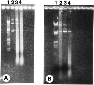
FIG. 2.
Photograph of isolated plasmids pIMP1eglATNF and pKNT19eglATNF after gel electrophoresis on a 1% agarose gel stained with ethidium bromide. (A) Lanes: 1, λ DNA digested with EcoRI and HindIII; 2, pIMP1eglATNF isolated from recombinant E. coli TG1; 3 and 4, pIMP1eglATNF isolated from recombinant C. acetobutylicum DSM792. (B) Lanes: 1, λ DNA digested with EcoRI and HindIII; 3, pKNT19eglATNF isolated from recombinant E. coli TG1; 2 and 4, pKNT19eglATNF isolated from recombinant C. acetobutylicum DSM792.

