Figure 5.
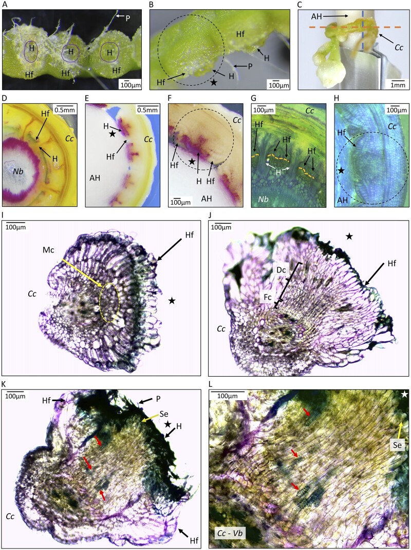
Anatomy of parasitic organs produced by C. campestris growing on an AH with MMS media with NAA and BA. A, Haustorial region inner face view. Haustoria (H) are observed in the center of the parasitic organ surrounded by the adhesion disk or holdfast (Hf). Upon removal from the AH, paper fibers (P) remain attached to the haustorial region. B, Lateral view of haustorial region with visible holdfast and protruding haustoria. Dotted line circle indicates a parasitic organ. C, Haustorial region attached to an AH. Orange dotted line indicates the sagittal plane of the haustorial region, sectioning direction used for (D–H). Dotted blue line indicates the transverse plane of the haustorial region, sectioning direction used for (I–L). D and G, Sections of C. campestris (Cc) growing on stems of N. benthamiana (Nb), with orange dotted lines in (G) indicating the epidermis of the host. E, F, and H, Cuscuta campestris growing on AHs. (D–F) were stained with phloroglucinol–HCl and fuchsia color indicates components of lignin. (G, H, and I–L) were stained with toluidine blue-O. H–L, Organized growth of haustorial cells on AH. H, Prehaustorial growth attached to AH, with star indicating outer layer of spindle. For (I–L), C. campestris was detached from the AH before sectioning, with the star indicating the face of C. campestris haustoria that had been in contact with the AH. I, Parasitic organ in an early developmental stage. Holdfast epidermal cells on the face were in contact with the host, so represent the adhesion disk. Yellow dotted line circles meristematic cells (Mc). J, A more developed parasitic organ showing File cells (Fc) and Digitate cells (Dc) positioned toward the side of the parasite facing the AH. K, Mature parasitic organ with elongated cells protruding through the middle of the Hf toward the AH. Searching hyphae are visible in the surface of the haustorium. L, Higher magnification of section of C. campestris in (K). Segments of tracheary elements (ring structures, indicated by arrows) are observed pointing in the direction of the haustorium tip that faces the AH. Sections in (E) and (F) were 20 dpi, (H) was 16 dpi and (I–L) were 36 dpi. Different stages of parasitic organ development were observed at 36 dpi in the same haustorial regions.

