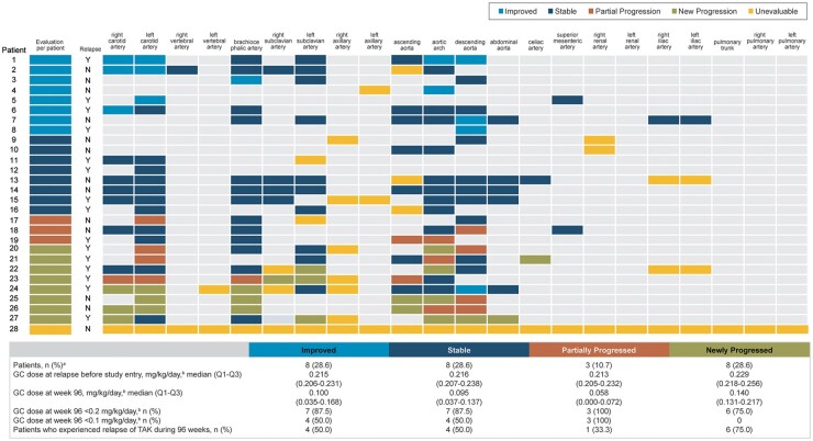
Fig. 4.
Change from baseline to week 96 in wall thickness and glucocorticoid doses (N = 28)
aOne patient was unevaluable for wall thickness. bPrednisone equivalent. Data are shown for all patients with central radiological assessment who received at least one dose of tocilizumab. Light grey spaces in the grid indicate no abnormal arteries were detected either at baseline or at week 96, and the patient’s condition was evaluated as stable. Imaging data were evaluated at 96 weeks (day 673; range, days 505–841). If multiple scans were available during the time window, the scan conducted closest to the scheduled date was used. Y: yes; N: no; GC: glucocorticoid; TAK: Takayasu arteritis.

