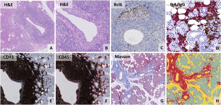
Fig. 1.
(Immuno-)histological analysis of parotid gland tissue from pSS patients
Parotid gland biopsies from pSS patients showing (A) a periductal focus on H&E staining with a centrally located lymphoepithelial lesion. (B) High-resolution image of the same lymphoepithelial lesion showing hyperplastic epithelium with intra-epithelial lymphocytes. (C) The presence of a germinal centre, as shown by a cluster of five or more Bcl6+ cells. (D) Dual staining for IgA (red) and IgG (brown) plasma cells showing an influx of IgG+ plasma cells. (E) CD45 staining of parotid gland tissue. (F) Digital image analysis of the relative area of CD45+ infiltrate by using QuPath. (G) Parotid gland tissue stained with a modified Masson stain in which dense connective tissue is coloured blue. (H) Digital image analysis of the modified Masson staining by using QuPath, in which red represents fibrotic tissue, yellow represents fat cells and green represents glandular parenchyma.

