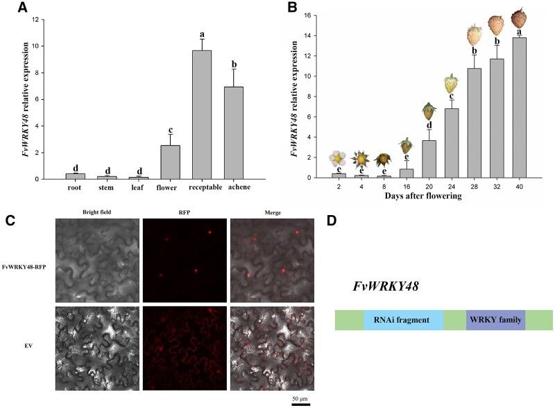
Figure 1.
Characterization of FvWRKY48 in wild strawberry. A and B, FvWRKY48 expression patterns in F. vesca in different organs and developmental stages. Expression levels represent the mean across three biological replicates, error bars represent standard deviation (sd) of mean, significant differences (P < 0.05) are indicated by lowercase letters based on Duncan’s test. C, Subcellular localization of the FvWRKY48–RFP fusion protein in N. benthamiana leaves. RFP alone was used as the control. Bar: 50 μm. D, The fragment for RNAi of FvWRKY48 gene. The green-colored boxes were CDS regions except RNAi fragment and WRKY family.

