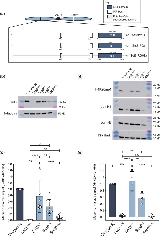
Fig. 4.
Set8(RG) mutant enzyme retains H4K20me1 in vivo. a) Diagram of Set8(WT), Set8(RG), and Set8(RGHL) proteins expressed from transgenes located on chromosome 3. b) Western blot of third instar larval brain extracts from Oregon-R wild-type and the indicated Set8 mutants using anti-Set8 and anti-β-tubulin antibodies. For uncropped membrane images, see Supplementary Fig. 5. c) Quantification of anti-Set8 signal on western blots by densitometry (see Materials and methods). Shown is the mean and standard deviation of measurements (circles) from technical replicates across four biological replicates. Oregon-R normalized signal was set to 1 for each replicate. Significance was determined by a one-way ANOVA followed by Tukey’s multiple comparison test. ** indicates P < 0.01, **** indicates P < 0.0001, and ns indicates not significant. d) Western blot of third instar larval nuclear extracts from Oregon-R wild-type and the indicated Set8 mutants using anti-H4K20me1, anti-pan H4, anti-pan H3, and anti-Fibrillarin antibodies. For uncropped membrane images see Supplementary Fig. 6. e) Quantification of anti-H4K20me1 signal on western blots by densitometry (see Materials and methods). Shown is the mean and standard deviation of measurements (circles) across three biological replicates normalized to pan H4 signal. Oregon-R normalized signal was set to 1 for each replicate. Significance was determined by a one-way ANOVA followed by Tukey’s multiple comparison test. ** indicates P < 0.01, **** indicates P < 0.0001, and ns indicates not significant.

