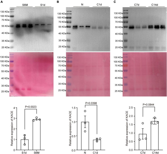
FIGURE 4.
Changes in protein pan-acetylation during sciatic nerve development, injury, and regeneration. Expression analysis of pan-acetylation during development (A), injury (B), and regeneration (C). The upper panel refers to Western blot results. The middle panel refers to Coomassie brilliant blue staining results that reflect the total amount of proteins on the PVDF membrane. The lower panel refers to statistical results. Bars represent the mean ± SEM, and the P-value is calculated by Student’s T-test, n = 3.

