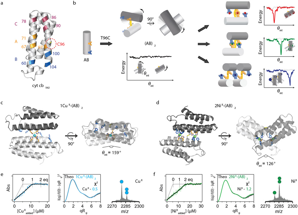
Figure 1 ∣. Design and characterization of the (AB)2 scaffold.
a, Locations of metal coordination motifs (A, B, C) and Cys96 on the cytochrome cb562 surface. b, Design of disulfide-linked (AB)2 with a malleable dimer interface, along with cartoon representations of possible metal-free/bound conformations and corresponding free-energy landscapes. Crystal structures of c, 1CuII-(AB)2 and d, 2NiII-(AB)2. CuII and NiII ions are represented as cyan and green spheres, respectively. X-ray data collection and refinement statistics are listed in Extended Data Table 2. Characterization of e, 1CuII-(AB)2 and f, 2NiII-(AB)2 complexes in solution using competitive metal titrations, solution SAXS, and ESI-MS. Log-scale SAXS plots are shown in Extended Data Figs. 2j-m. Changes in Fura-2 absorbance at 335 nm (left) are plotted with theoretical metal-binding isotherms (grey) in the absence of (AB)2. Experimental data points and error bars are presented as mean and standard deviation of three independent measurements. Theoretical SAXS profiles (middle) derived from the crystal structures of 1CuII-(AB)2 (cyan) and 2NiII-(AB)2 (green) are plotted with experimental SAXS profiles (black) and corresponding χ2 values. Circles in ESI-MS spectra (right) represent the number of NiII (green) and CuII (cyan) ions bound to (AB)2.

