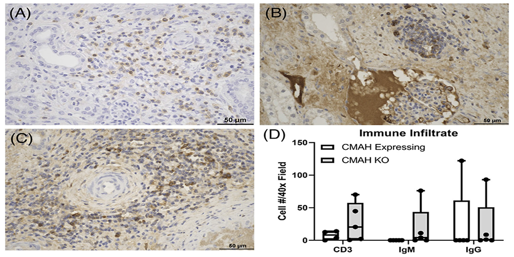
FIGURE 4.

Increased T and B cell infiltrates in the renal interstitium of CMAHKO kidney transplants. (A-C) Representative images of (A) CD3, (B) IgM, and (C) IgG from B1417 (Group 2). The majority of cells reside around medium caliber arteries and within the renal interstitium. (D) Numbers of CD3+ T cells, IgM+ B cells, and IgG+ B cells in CMAHKO pig kidney transplants. No statistically significant differences were noted despite trends for increased T and B cell infiltrates in some of the CMAHKO kidneys. Immunohistologic staining (brown color). Variance in each group is expressed as standard deviation (n = 4 data points/group). (Size bar 50 μm)
