BACKGROUND
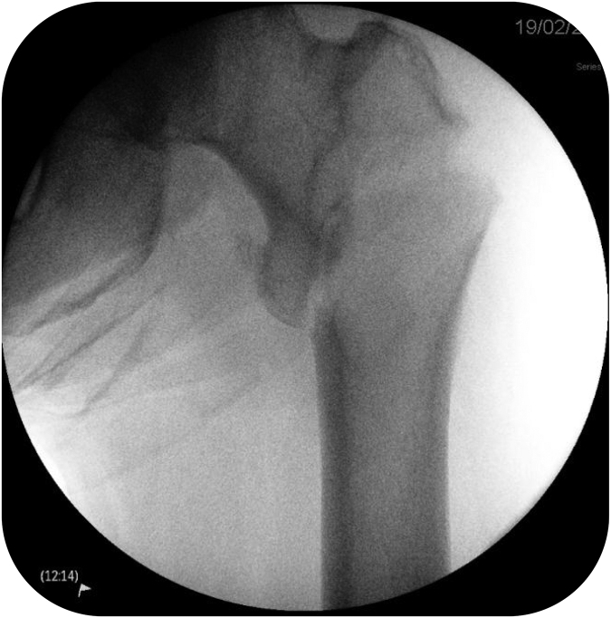
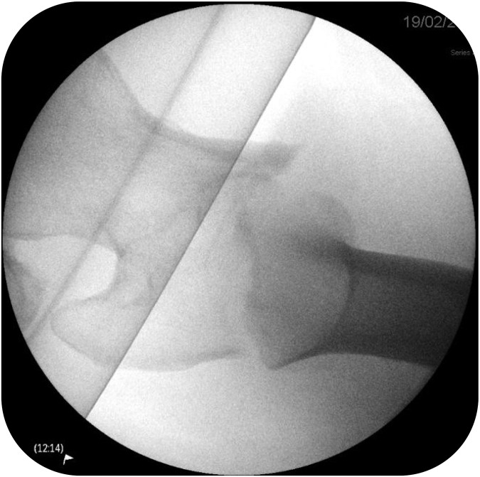
Although common, neck of femur (NOF) fractures requiring surgical fixation can be difficult to manage.1,2 This can be particularly challenging when the lesser trochanter (LT) and greater trochanter (GT) are both attached to the proximal fragment due to the resultant pull of muscles (Figures 1 and 2). Fragment-specific fixation allows reduction to be maintained while definitive cephalomedullary fixation is introduced.
Figure 1 .
Anteroposterior image of fracture (after attempted closed reduction)
Figure 2 .
Lateral of fracture (after attempted closed reduction)
TECHNIQUE
Fragment-specific reduction techniques can be employed:
-
1.
Reduce the LT with large bone-holding forceps.
-
2.
Reduce the GT using pointed reduction forceps.
-
3.
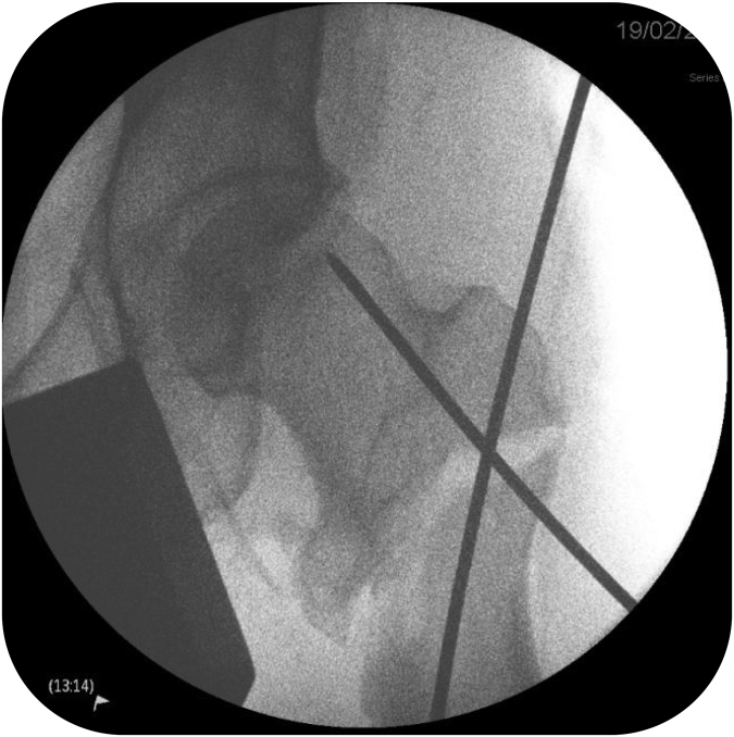
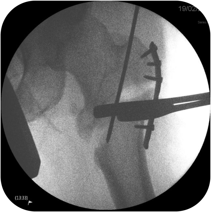
Hold reduction using 2mm crossed Kirschner wires (Figure 3).
-
4.
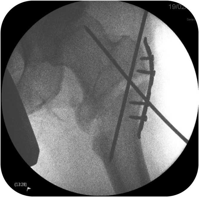
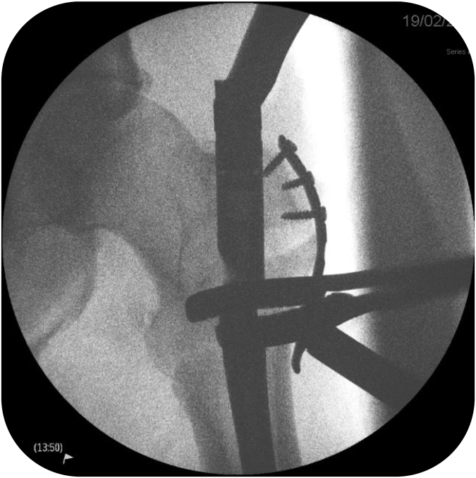
Apply small fragment-locking plate (EVOS plate, Smith+Nephew, Croxley Park, UK) and secure with unicortical locking screws to neutralise the abduction forces (Figure 4). Plan placement so as to avoid the entry point for the neck screw.
-
5.
Apply large bone-holding forceps to the LT to reinforce the reduction (Figure 5).
-
6.
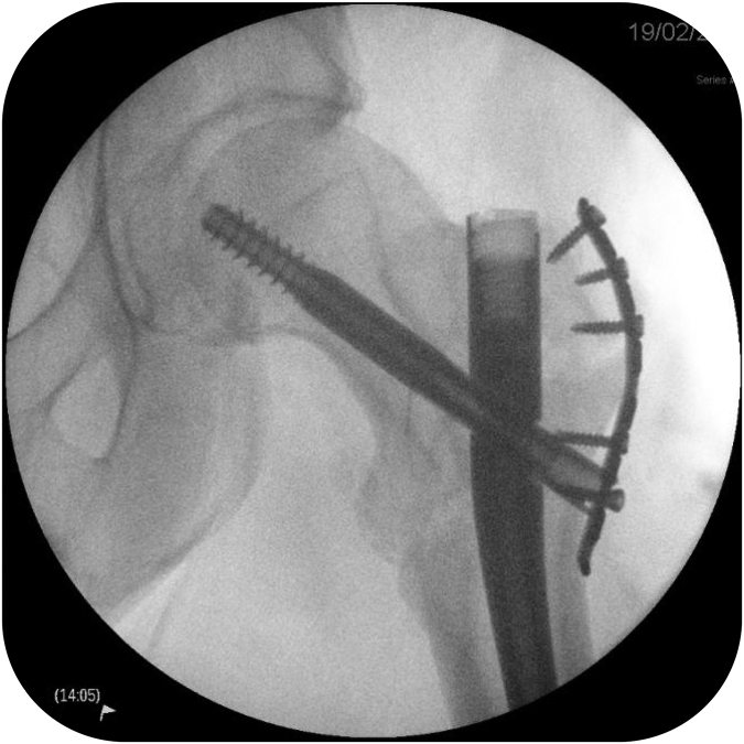
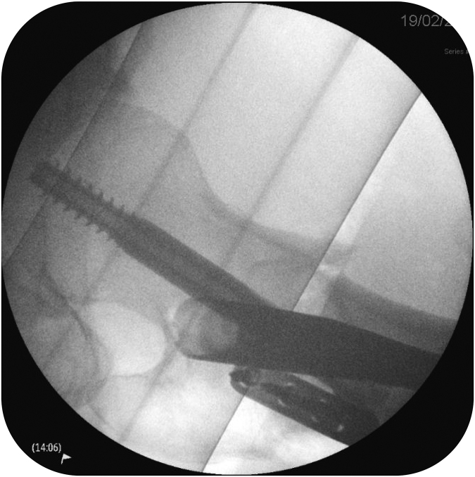
Medialise the entry point for a trochanteric entry cephalomedullary nail (Gamma3, Stryker, Newbury, UK) as described by Westacott and Bhattacharava3 (Figure 6).
Figure 3 .
Crossed Kirschner wires
Figure 4 .
EVOS plating
Figure 5 .
Addition of Hey-Groves bone-holding forceps before intramedullary nailing
Figure 6 .
Intramedullary nail insertion
DISCUSSION
Anatomical reduction of NOF fractures can be challenging. Fracture-specific reduction can be used to stabilise the proximal femur to allow definitive fixation, and to avoid varus reduction of unstable NOF fractures (Figures 7 and 8).
Figure 7 .
Final anteroposterior image
Figure 8 .
Final lateral image
References
- 1.National Institute for Health and Care Excellence (NICE). Clinical Guideline 124 (CG124). Hip Fracture: Management. https://www.nice.org.uk/guidance/cg124 (Updated May 2017). [PubMed]
- 2.Wang ZH, Li KN, Lan Het al. . A comparative study of intramedullary nail strengthened with auxiliary locking plate or steel wire in the treatment of unstable trochanteric fracture of femur. Orthop Surg 2020; 12: 108–115. [DOI] [PMC free article] [PubMed] [Google Scholar]
- 3.Westacott DJ, Bhattacharava S. A simple technique to help avoid varus malreduction of reverse oblique proximal femoral fractures. Ann R Coll Surg Engl 2013; 95: 74. [DOI] [PMC free article] [PubMed] [Google Scholar]


