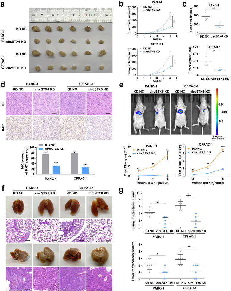
Fig. 3.
CircSTX6 facilitates the tumorigenesis and metastasis of PDAC cells in vivo. a Representative picture of subcutaneous xenograft tumors (n = 6 for each group). b-c Curves of tumor volumes and weights show negative effects of circSTX6 knockdown on the formation of subcutaneous xenograft tumors. d HE and Ki-67 IHC staining of xenograft tumors. Original magnification 400×. Scale bar = 50 μm. H-scores of the Ki67 staining. e Representative images and analysis of luminescence intensity in tail vein tumor metastasis mouse models (n = 6 for each group). f Representative images and HE staining of metastatic nodules in the lungs and livers of mice. Original magnification 100×. Scale bar = 200 μm. g The numbers of lung and liver metastatic nodules were measured. (Values are expressed as the means ± SDs; *P < 0.05, **P < 0.01 and ***P < 0.001)

