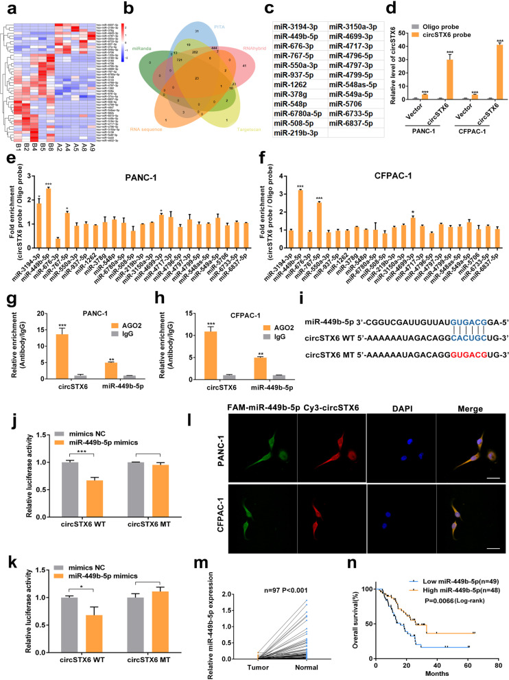
Fig. 4.
CircSTX6 serves as a sponge for miR-449b-5p. a Clustered heatmap of the differentially expressed miRNAs in PDAC tissues from five T4 PDAC patients with long survival time and tissues from five T4 PDAC patients with short survival time. b Venn diagram showing the overlap of the target miRNAs of circSTX6 predicted by miRanda, PITA, RNAhybrid, TargetScan and the results of RNA-seq. c. Table of target miRNAs selected for circSTX6. d The efficiency of the circSTX6 probe in PDAC cells was validated using qRT–PCR after the RNA pull-down assay. A random oligo probe served as a negative control. e-f The expression levels of 23 miRNA candidates were detected in the RNAs pulled down by circSTX6 and oligo probes. g-h Anti-AGO2 RIP was performed to detect circSTX6 and miR-449b-5p in PDAC cells. i A schematic of the wild-type (WT) and mutant (MUT) circSTX6 luciferase reporter vectors. j-k A luciferase reporter assay was used to confirm the interaction between circSTX6 and miR-449b-5p. l The colocalization of circSTX6 and miR-449b-5p in PDAC cells was detected using a FISH assay. The circSTX6 probe was labeled with Cy3 (red), the miR-449b-5p probe was labeled with FAM (green), and nuclei were stained with DAPI (blue). Original magnification 400 ×. Scale bar = 50 μm. m The expression of miR-449b-5p was detected in 97 PDAC tissues and corresponding noncancerous tissues. n Kaplan–Meier analysis of miR-449b-5p expression and overall survival in 97 patients with PDAC. The median expression value of miR-449b-5p was used as the cutoff (n = 49). Log-rank tests were used to determine the statistical significance. (Values are expressed as the means ± SDs; *P < 0.05, **P < 0.01 and ***P < 0.001)

