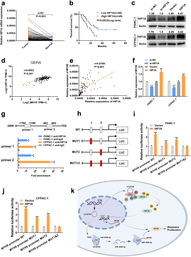
Fig. 9.
HIF1A promotes MYH9 transcription. a-b. The expression level of HIF1A in PDAC tissues and matched noncancerous tissues was detected by qRT–PCR. Kaplan–Meier analysis was used to illuminate the correlation between the level of HIF1A and the survival time of PDAC patients. The median expression value of HIF1A was used as the cutoff (n = 49). Log-rank tests were used to determine the statistical significance. c. HIF1A knockdown and overexpression cell lines were constructed. d. GEPIA analysis showed a positive relationship between MYH9 and HIF1A in PDAC tissues. e. Correlation analysis showed a positive relationship between MYH9 and HIF1A in 97 PDAC tissues. 18 S served as the internal control. f. MYH9 mRNA expression was significantly downregulated in HIF1A-knockdown PDAC cells and upregulated in HIF1A-overexpressing PDAC cells. g. CHIP assays revealed that HIF1A could bind to two fragments (-1194 – -1185, -561 – -552) of the MYH9 promoter. h-j. Luciferase reporter assays were used to confirm the interaction between HIF1A and two fragments (-1194 – -1185, -561 – -552) of the MYH9 promoter. k. Schematic diagram illustrating the mechanism by which circSTX6 promotes PDAC proliferation and metastasis through posttranscriptional and transcriptional regulation of MYH9 by sponging miR-449b-5p and inhibiting VHL-EloBC-CUL2-Rbx1 complex-dependent ubiquitination of HIF1A, respectively. (Values are expressed as the means ± SDs; *P < 0.05, **P < 0.01 and ***P < 0.001)

