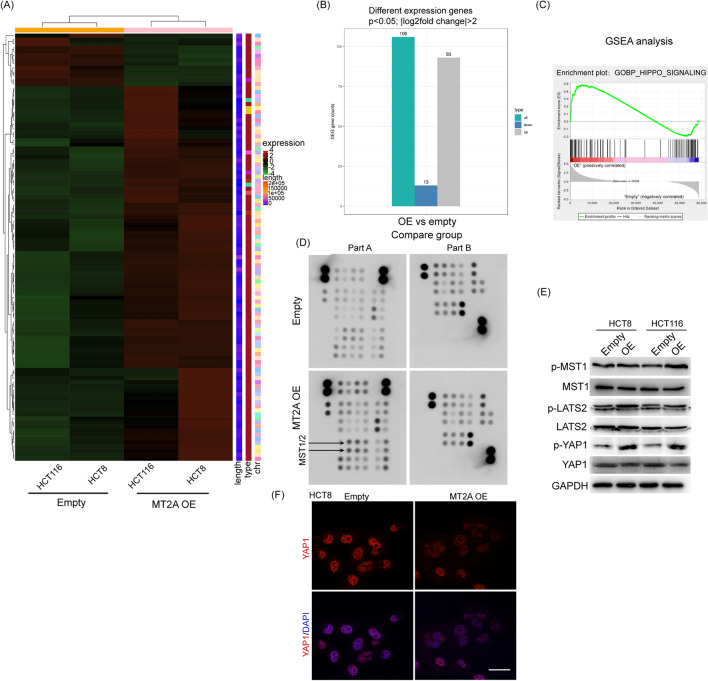
Fig. 5.
MT2A promotes Hippo signaling in CRC cells. A RNA sequencing was used to evaluate the gene expression level between MT2A-overexpressing and control HCT8 and HCT116 cells. B With a cutoff of p < 0.05 and |log2fold change|> 2, 106 genes were selected after MT2A overexpression in both cell lines, including 93 upregulated genes and 13 downregulated genes. C GSEA showed that MT2A overexpression significantly enriched Hippo signaling in HCT8 and HCT116 cells (normal p value < 0.05, NES > 1.0). D We used a Proteome Profiler Array Human Phospho-Kinase Array to identify the key variations in phosphokinases after transfection of MT2A. This array is a rapid and sensitive product used to detect relative levels of phosphorylation of 43 kinase phosphorylation sites and GAPDH protein. The A-B5 and B6 sites represent the expression levels of p-MST1 and p-MST2, respectively. MT2A overexpression promoted the phosphorylation of MST1 and MST2. E In HCT8 and HCT116 cells, the expression levels of p-MST1, MST1, p-LATS2, LATS2, p-YAP1, and YAP1 were determined by Western blot analysis. MT2A overexpression increased the phosphorylation of p-MST1, p-LATS2 and p-YAP1 in both cell lines. F Immunofluorescence staining of YAP1 showed that MT2A overexpression inhibited nucleus location of YAP1 in HCT8 cells. OE overexpression. Scale bar = 5 μm

