FIGURE 1.

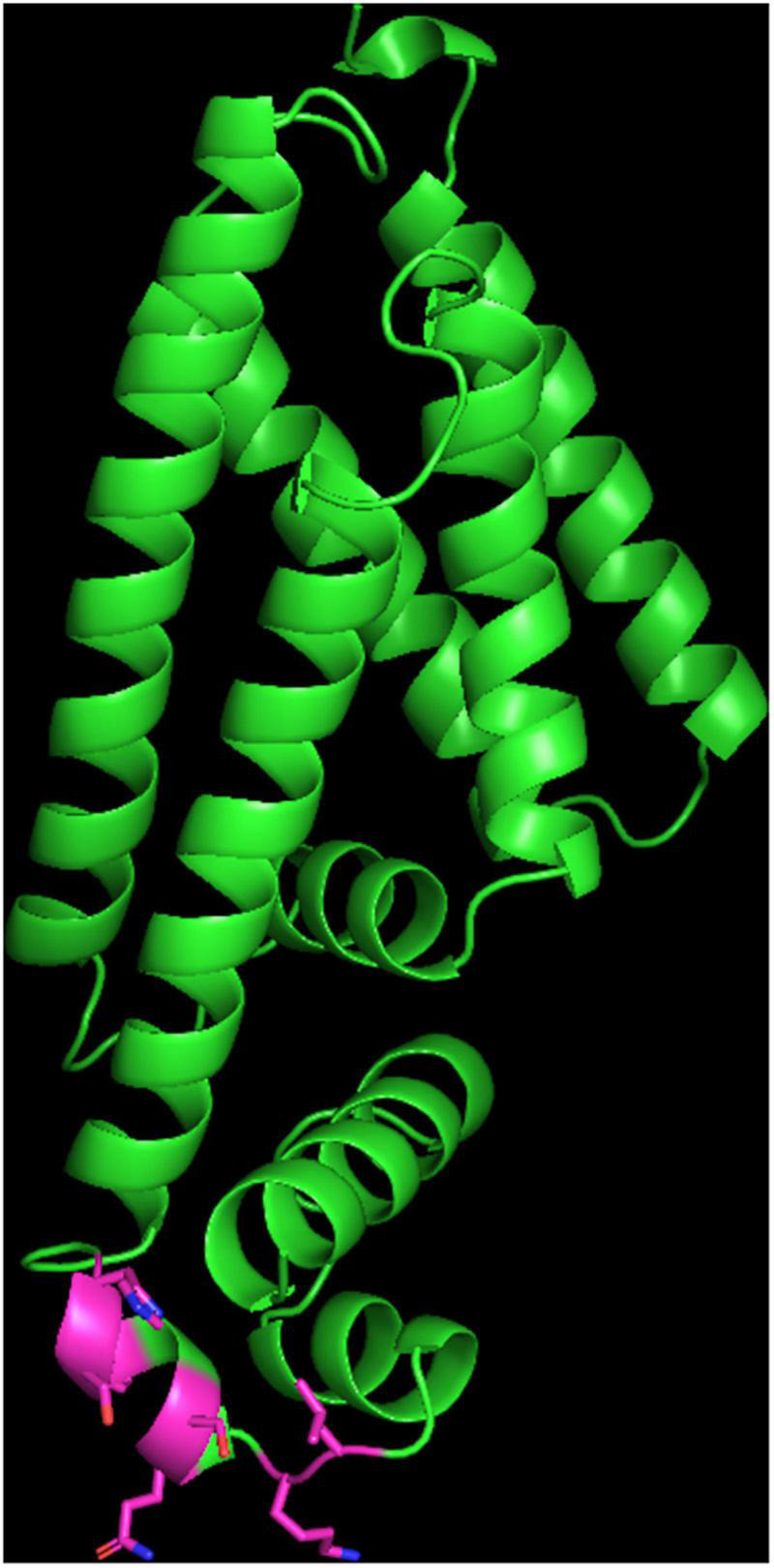
Structure of the FepR protein. The carbon of the side chain of the mutated residues inside the HTH domain are shown in pink. Oxygen atoms are shown in red and nitrogen in blue. Hydrogen atoms are hidden.

Structure of the FepR protein. The carbon of the side chain of the mutated residues inside the HTH domain are shown in pink. Oxygen atoms are shown in red and nitrogen in blue. Hydrogen atoms are hidden.